Fig. 7.
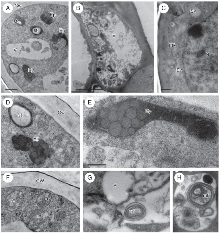
Zygostates lunata, TEM. (A) Transverse section of apical part of secretory hair showing elaioplasts, plastids containing starch, vacuoles and granular cytoplasm. (B) Basal part of secretory hair with parietal cytoplasm containing numerous plastids and abundant ER profiles. (C) Parietal cytoplasm of hair with ER, secretory vesicles and nucleus. (D) Detail of (A) showing two kinds of plastid: the first almost completely occupied by a starch grain, the other, a typical elaioplast containing numerous lipid droplets. (E) Detail of elongate elaioplast containing numerous lipid droplets. (F) ER membranes and mitochondria in parietal cytoplasm of secretory hair. (G) Vacuoles containing membranes, small vesicles and myelin-like figures. (H) Vacuole enclosing numerous myelin-like figures. Scale bars: (A, D, G, H) = 1 µm; (B) = 50 µm; (C, E, F) = 0·50 µm. See Fig. 1 for abbreviations.

