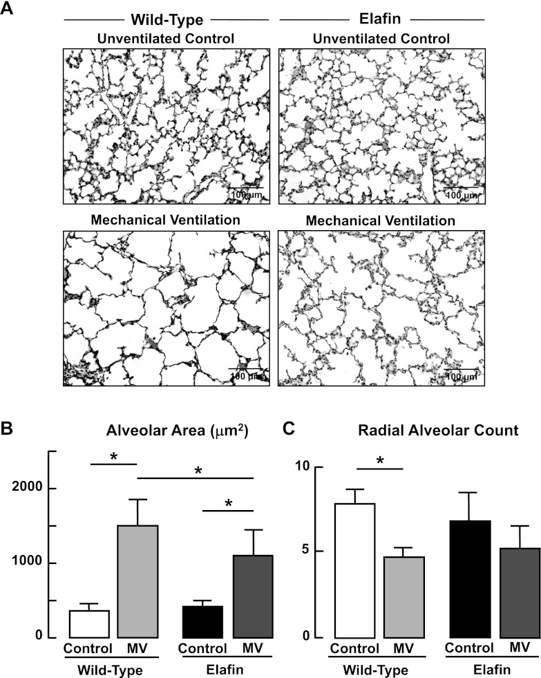
Fig. 10.
Impaired alveolar formation elicited by MV-O2 is attenuated in elafin-expressing mice. A: representative lung tissue sections (×200) from 6-day-old wild-type and elafin-expressing mice after MV-O2 for 36 h, showing increased air space size in both groups when compared with unventilated controls that breathed 40% O2 for 36 h. B: summary data (means and SD) for alveolar area, assessed by quantitative image analysis of lung tissue sections from wild-type and elafin-expressing mice after MV-O2 for 36 h, compared with unventilated controls. C: summary data (means and SD) for radial alveolar counts, an index of alveolar number, of lung tissue sections from wild-type and elafin-expressing mice after MV-O2 for 36 h, compared with unventilated controls that breathed 40% O2 for 36 h. Radial alveolar counts decreased significantly after MV-O2 of wild-type mice, whereas there was not a significant reduction seen in the lungs of elafin-expressing mice. *Significant difference between groups, P < 0.05; n = 4–5/group.

