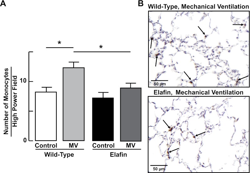
Fig. 7.
Lung influx of monocytes during MV-O2 is suppressed in elafin-expressing mice. A: summary data (means and SD) showing increased numbers of monocytes (counted in 20 ×400 fields) in lungs of 6-day-old wild-type mice after MV-O2 for 24 h, compared with unventilated controls that breathed 40% O2 for 24 h. Monocyte counts did not increase significantly in response to MV-O2 in elafin-expressing mice compared with unventilated controls. *Significant difference between groups, P < 0.05. Values are means and SD; n = 4–6/group. B: representative images of lung tissue sections (×400) showing monocytes (arrows, brown stain of F4/80 monocyte antibody) in lungs of wild-type (top) and elafin-expressing (bottom) mice after MV-O2 for 24 h.

