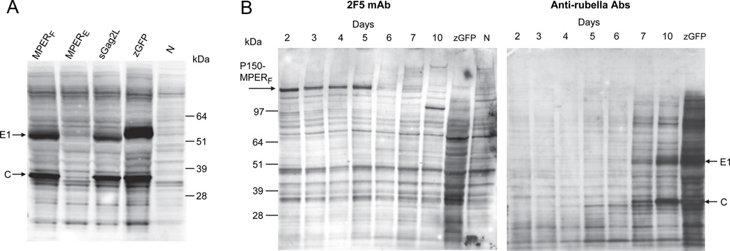
Figure 2. Vector replication and expression of HIV MPER determinants.
(A) Rubella vector replication at passage P0 was detected by Western blot with antibodies specific for rubella structural proteins C (33 kDa) and E1 (58 kDa). Strong vector replication was observed for the MPERF vector and for sGag2L, but not for MPERE (all in the vaccine strain). These were compared to a control vector expressing zGFP in wild type rubella or to uninfected cells (lane N). (B) Time course of MPERF expression (at passage P3), as detected by Western blot with monoclonal 2F5 (left panel) or with antibodies to rubella structural proteins (right panel). MPERF was expressed as a high molecular-weight fusion protein with P150 (arrow). Maximal expression was observed from day 2 to 5 after infection. In contrast, rubella structural proteins E1 and C first appeared on day 5 or 6 and were strongly expressed on days 7 to 10. The same samples were used for both gels.

