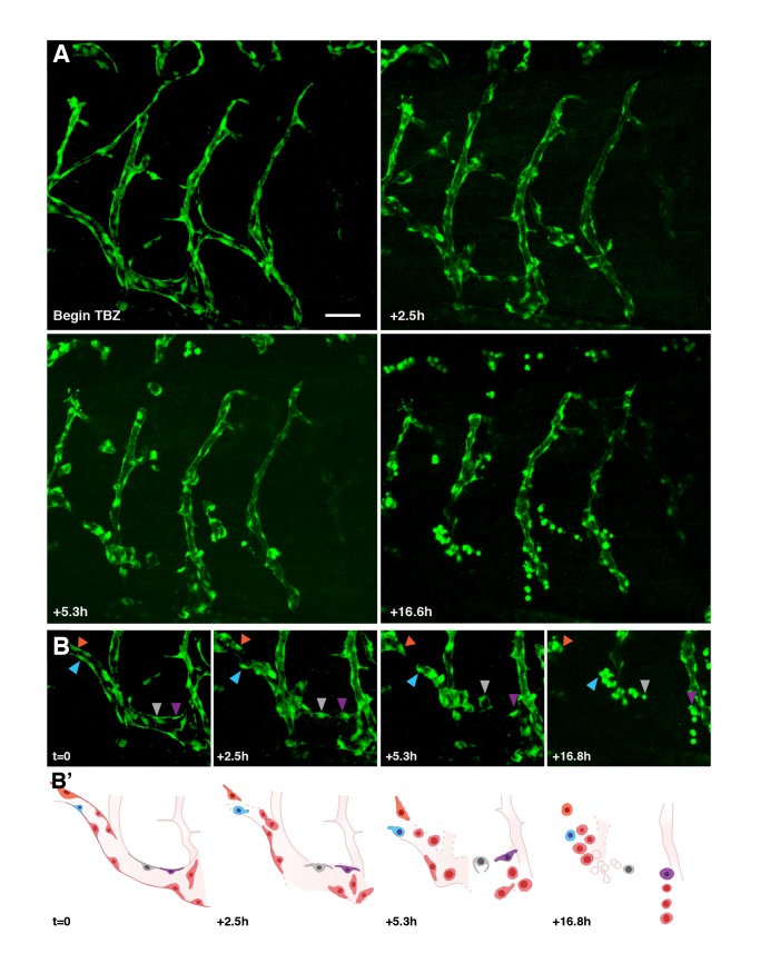
Figure 5. TBZ disrupts newly established vasculature, as visualized in vivo using time-lapse fluorescence microscopy within kdr:GFP frogs.
Retraction and rounding of vascular endothelial cells (arrowheads) is apparent in TBZ-treated embryos (A, time lapse of frogs treated as in Figure 3) as compared with continued vascular growth in control animals (Figure S7). Scale bar, 80 µm. (B) A series with intermediate time points is shown for a sub-region of that shown in (A). (B′) Schematics of the images in (B) indicate positions of specific cells.

