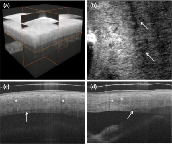
Fig. 3.
In vivo 3-D imaging of human limbus. (a) rendering and 3-D reconstruction of data set. (b) C-scan (x-y cross section). Arrows show Schlemm's canal. (c) B-scan in fast scanning axis (y-z cross-section). (d) B-scan in slow scanning axis (x-z cross section). Protocol C from Table 1 was used for scanning the eye. Asterisks show blood vessel shadows.

