
Fig. 2.
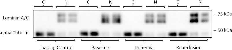
Cytoplasmic and nuclear separation. Representative blots showing adequate separation of cytoplasmic and nuclear fractions were assessed via Western blot analysis detection of α-tubulin, cytoplasmic protein (C), and laminin A/C, nuclear protein (N), in each lane. A single muscle homogenate sample was loaded in duplicate with each gel and used as a between-blot internal loading control. All gels were run in with cytoplasmic and nuclear fractions loaded in adjacent cells, i.e., baseline cytoplasmic next to the baseline nuclear fraction as shown in the representative blot.