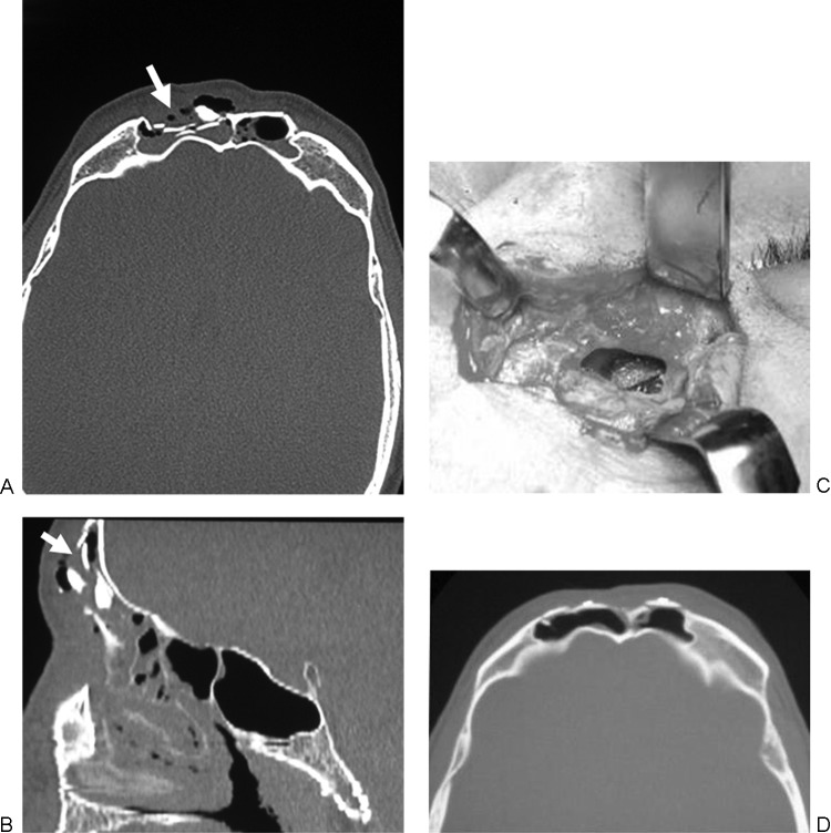
Figure 2.
Type A—Illustrative case: a 43-year-old man with fracture of the anterior wall of the frontal sinus. (a, b) Axial computed tomography (CT) scan and sagittal CT reconstruction show fracture of the anterior wall of the frontal sinus (white arrows) without evolvement of the posterior wall; (c) intraoperative findings: a bone graft taken from the mandible was placed to repair the fracture; (d) postoperative CT scan showing surgical repair of the anterior wall of the frontal sinus.

