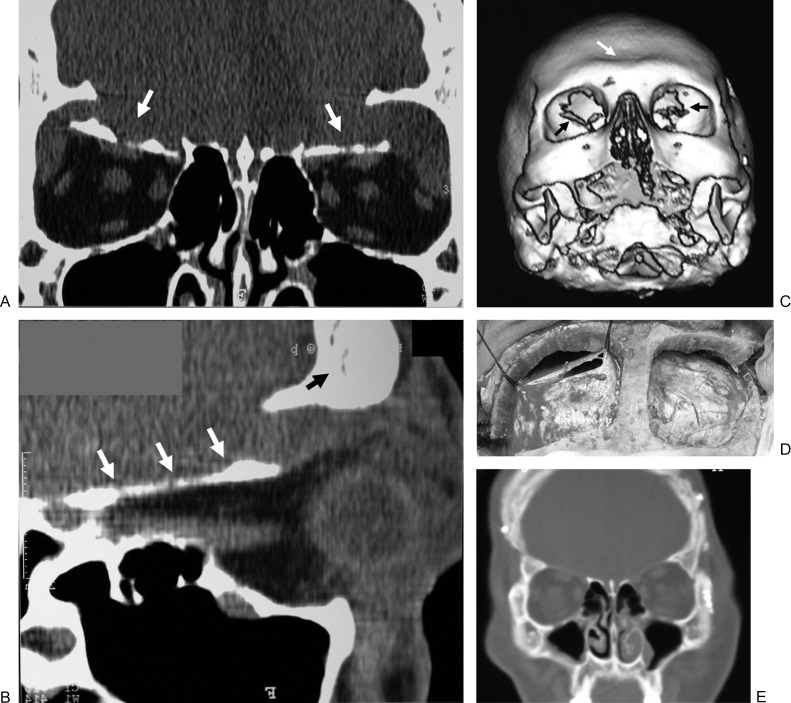
Figure 4.
Type C—Illustrative case: a 22-year-old man with bilateral fractures of the orbital roof and a little parietal fracture. (a) Coronal computed tomography (CT) scan showing bilateral depression of the orbital roof (white arrows); (b) sagittal CT reconstruction showing fracture of the orbital roof (white arrows) without evolvement of the frontal sinus (black arrow); (c) three-dimensional reconstruction showing bilateral dislocation of the orbital roof (black arrows) without fracture of the anterior wall of the frontal sinus (white arrow); (d) intraoperative field: bone fragments have been removed, dural laceration was repaired and suspended; (e) postoperative CT scan.

