Fig. 1.
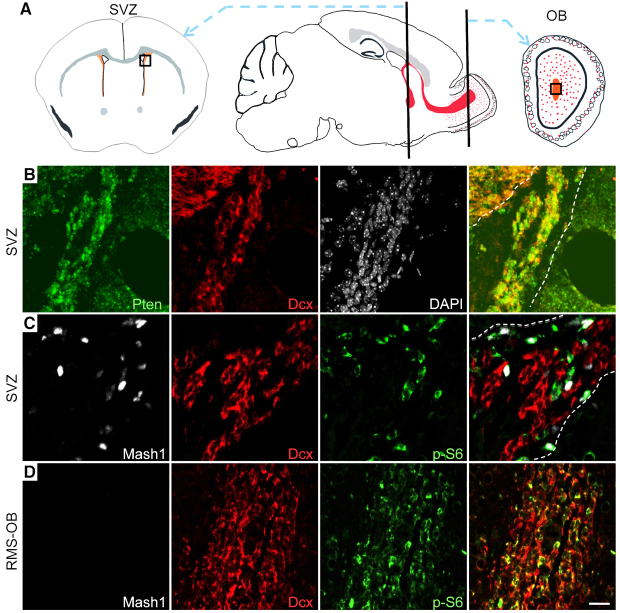
The PI3K-mTorc1 pathway was inactive in migrating SVZ neuroblasts, but activated in OB neuroblasts. (A) Schematic brain sections indicating anatomical regions in the SVZ and OB (black boxes) from which images were captured. (B-D) IF staining of the SVZ (B,C) and the terminal RMS at the OB core (D) in coronal sections from 6-month-old (B,D) or 18-day-old (C) wild-type mice. (B) Pten (green) and Dcx (red) double IF labeling of the SVZ. Nuclei were labeled with DAPI (white). Pten was expressed in Dcx+ cells in the SVZ (overlay of Pten and Dcx, far right panel). (C,D) Mash1 (white), Dcx (red) and p-S6 (green) triple IF labeling of the SVZ (C) and OB (D) (overlay, far right panel). p-S6 was expressed in Mash1+ transit-amplifying cells but not Dcx+ neuroblasts in the SVZ (C), whereas in the OB, a substantial population of Dcx+ neuroblasts expressed p-S6 (D). The white dashed lines in the overlay mark the boundary of the SVZ in panels A and B. Scale bar: 20 μm.

