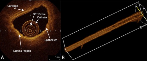
Fig. 5.
Optical coherence tomography (OCT) images of a medium-sized airway in cross-section (A) and reconstructed 3-dimensional airway obtained by a “pull back technique” where the OCT probe is retracted 5 cm up the airway during scanning (B). OCT probe and surrounding catheter can be seen in lumen of A. A also shows the bright contrast pattern of the lamina propria and the less contrast of the epithelial layer and the cartilage. The pull back technique produces images of an airway path while the OCT probe is automatically retracted 5 cm. Images are obtained in a “helical” method and can be reconstructed into the 3-dimensional image of the airway path. (Images courtesy of Dr. Keishi Ohtani, BC Cancer Research Centre, Vancouver, BC, and Department of Surgery, Tokyo Medical University, Tokyo, Japan).

