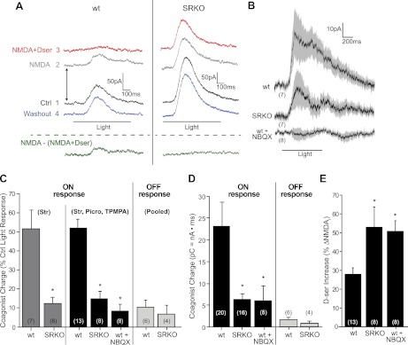
Fig. 3.
Light-evoked coagonist release is diminished in SRKOs and blocked by NBQX. RGC responses were recorded at +40 mV. Recordings were made in 1 mM Mg2+, 1 μM TTX, 10 μM strychnine, 50 μM TPMPA, and 50 μM picrotoxinin. TPMPA and picrotoxinin were excluded where specified. A: raw trace showing the effects of bath-applied NMDA (100 μM) or NMDA + d-serine (100 μM) on light-evoked ON responses. Numbers indicate the order of drug application. Traces are offset to show the baseline shift in current caused by the different pharmacological conditions (arrows). The green trace, representing coagonist release, was obtained by subtracting the NMDA + d-serine trace from the NMDA trace. B: average coagonist release traces (SE shown in gray) for ON cells compared between wt and SRKO RGCs in control conditions and wt RGCs in the continuous presence of NBQX. C: charge carried by coagonist release in the resultant trace normalized to the charge of the initial light response. ON responses were significantly larger in wt than SRKO either when only strychnine (Str) was present or when TPMPA and picrotoxinin (Picro) were also present. Coagonist release was lower for both genotypes during OFF responses compared with ON responses. No difference between genotypes was observed for OFF responses (data pooled for just strychnine and strychnine, TPMPA, and picrotoxinin). NBQX significantly reduced coagonist release during ON responses in wt. D: total charge (picocoulombs) carried by coagonist release. Pooled data with and without GABAergic block. E: % increase in the baseline shift (see A) from NMDA after adding NMDA + d-serine. d-Serine-induced potentiation of NMDAR currents (ΔNMDAi) was larger in SRKO than in wt. In the continuous presence of NBQX, d-serine potentiation was enhanced in wt. Number of cells recorded from (n) indicated in parentheses. *P < 0.05 compared with wt in control conditions.

