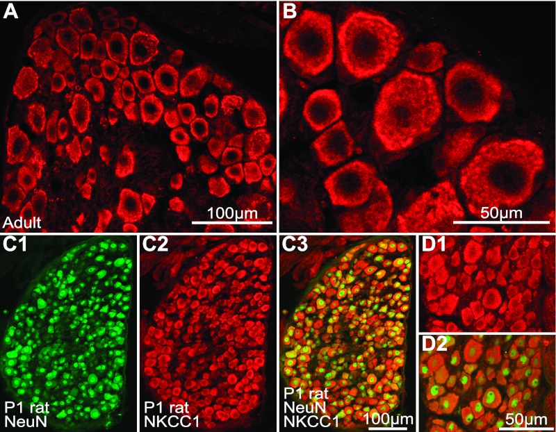
Fig. 3.
NKCC1 immunolabeling in adult and newborn rat DRG neurons. A and B: adult rat DRG section at low (A) and high (B) magnification showing NKCC1-IR (Cy3, red, Kaplan-CT antibody) in large and small neurons. NKCC1-IR was localized toward the cell periphery as in mouse DRG (Fig. 2D) but was also observed in the cytosol. C: newborn (P1) rat DRG section dual-immunolabeled for NeuN (FITC, green, C1) and NKCC1 (Cy3, red, C2). C1: NeuN immunofluorescence alone. C2: NKCC1 immunofluorescence alone. C3: superimposition of NeuN and NKCC1 immunofluorescence. D1: high magnification of NKCC immunofluorescence shown in C2. D2: merged NKCC1 and NeuN immunofluorescence. NeuN-IR is higher in the cell nucleus; NKCC1-IR is observed in all DRG neurons, and it is characteristically distributed throughout the cytosol in newborn rat.

