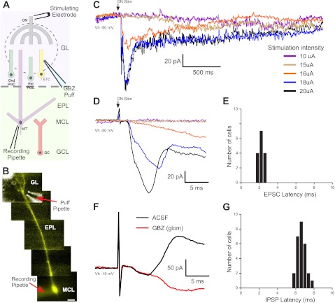
Fig. 1.
Latencies of mitral cell (MC) excitatory and inhibitory responses to olfactory nerve (ON) stimulation. A: schematic of the experimental setup indicating the location of the pipette used to puff gabazine (GBZ). GL, glomerular layer; ONd PGC, ON-driven periglomerular cell (PGC); ETd PGC, external tufted cell (ETC)-driven PGC; EPL, external plexiform layer; MCL, MC layer; GCL, granule cell (GC) layer. B: MC showing a Lucifer yellow (LY) dye-filled apical dendrite, superimposed with a bright field image of the GBZ puff pipette. C: MC responses to ON stimulation with increasing stimulation strength [holding potential (Vh): −50 mV, average of 6–8 sweeps]. In this cell, stimulation intensities below 16 μA elicited no response (purple trace: 10 μA, brown trace: 15 μA); at 16 μA, the cell responded with a slow excitatory ramp into a long-lasting depolarization (LLD; red trace). At 18 μA or greater, a short-latency excitatory postsynaptic current (EPSC) and LLD occurred (blue trace: 18 μA, black trace: 20 μA). D: expanded traces from A showing the latency and onset of ON-evoked MC responses. MCs held at −50 mV responded to suprathreshold ON stimulation with a short-latency EPSC (downward deflection) followed by an inhibitory current (upward deflection). E: histogram showing EPSC latency to ON stimulation in 15 MCs. F: in artificial cerebrospinal fluid (aCSF; black trace, average of 10 sweeps), MCs held at −10 mV showed only the prominent upward-deflection inhibitory postsynaptic current (IPSC). Microinjection of 100 μM GBZ into the glomerulus containing the MC apical dendrite abolished the IPSC (red trace, average of 10 sweeps). G: histogram of MC IPSC latencies to ON stimulation in aCSF (n = 27, solid bars).

