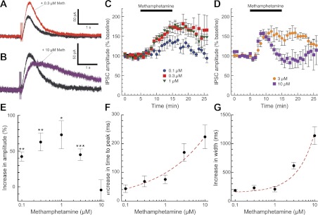
Fig. 1.
Methamphetamine has bidirectional, concentration-dependent effects on dopamine inhibitory postsynaptic current (IPSC) amplitudes. We obtained whole cell patch-clamp recordings of dopamine-mediated IPSCs in substantia nigra and ventral tegmental area (VTA) dopamine neurons (Beckstead et al. 2004) in the presence of GABA, glutamate, and nicotinic acetylcholine receptor blockers. Bath perfusion of a low concentration of methamphetamine (0.3 μM, red trace) significantly enhanced the amplitude and slightly prolonged the duration of dopamine IPSCs (A and C; n = 6–13 cells from 3–9 mice). Perfusion of a high concentration of methamphetamine (10 μM, purple trace) briefly enhanced IPSC amplitudes but subsequently decreased IPSC amplitudes to baseline levels or slightly below (B and D; n = 6–12 cells from 3–6 mice). Thus methamphetamine exhibited an inverted U-shaped concentration-effect curve on dopamine IPSC amplitudes, determined as the average amplitude 10–12 min after the beginning of methamphetamine perfusion (E; paired t-tests for each concentration: *P < 0.05, **P < 0.01, ***P < 0.001). Methamphetamine effects on IPSC kinetics were examined by measuring time to peak (F) and width at 50% maximum amplitude (G). The slowing of IPSC kinetics was progressively enhanced by higher methamphetamine concentrations and was modeled well by a single exponential increase (red dashed lines).

