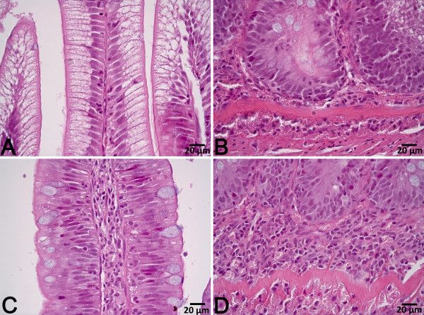
Figure 2.
Representative images of distal intestine tissue from fish fed diet PPC (A & B) and fish fed diet PPC + S (C & D). The tissue from PPC + S fed fish exhibited reduced enterocyte vacuolization and abnormal nucleus position, increased lamina propria and submucosa width with prominent leukocyte infiltration.

