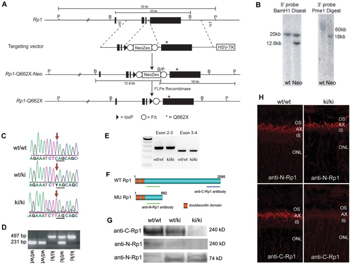
Figure 2. Generation and Characterization of Rp1 Q662X/Q662X Knock-in Mice. A.
Gene targeting strategy to introduce Q662X mutation into the endogenous Rp1 locus. The gene targeting vector was produced by introducing the Neo-Zeo selection cassette and Q662X mutation into the Rp1 gene in a BAC which contains 140 kb of mouse genomic DNA including the Rp1 locus, using homologous recombination techniques. The gene targeting vector was then retrieved from the modified BAC, and used to transfect mouse ES cells. Correctly targeted ES cells were injected into blastocysts to generate Rp1-Neo-Q662X mice. Crosses to Flpe deleter mice were used to remove the Frt-flanked Neo-Zeo selection cassette and generate the final Rp1-Q662X allele. B, BamHI restriction sites; P, PmeI restriction sites. B. Southern blots using the 5′ and 3′ probes indicated in A demonstrating correct targeting of the Rp1 locus in mouse ES cells. The 12.6 and 18 kb bands in the 5′ and 3′ blots, respectively, indicate correct recombination to generate the Rp1-Neo-Q662X allele. The large size of the wild-type Pme1 restriction fragment (60 kb) in the 3′ blot makes it difficult to resolve this band with standard electrophoresis techniques. C. Sequence traces from PCR products amplified from wild-type, heterozygous (wt/ki) and homozygous (ki/ki) mice. D. Example PCR-based genotyping of Rp1-Q662X (ki) knock-in mice. Using PCR primers that amplify across the residual Frt-LoxP sites, the wild-type and ki alleles can be readily distinguished. E. RT-PCR of retinal RNA from mice of indicated genotypes showing the correct splicing of mutant Rp1-Q662X allele as that of wild type Rp1 allele, using primer sets bridging exon 2 to 3 and exon 3 to 4. F. Predicted proteins produced by the wild-type (wt) and knock-in (ki) Rp1 alleles, showing locations of the anti-N-Rp1 and anti-C-Rp1 antibodies used in panels G and H. G. Western blot of retinal extracts from mice of the indicated genotypes showing production of the 74 kD truncated protein by the knock-in (ki) allele. Note that there is no full-length Rp1 protein detected in the retinas of the homozygous ki/ki mice by either the anti-N-Rp1 or anti-C-Rp1 antibodies. H. Immunofluorescence analysis of Rp1 proteins (red) produced in the retinas of wild-type (wt/wt) vs. homozygous knock-in (ki/ki) mice using the anti-N-Rp1 and anti-C-Rp1 antibodies indicated in panel E. Note that the truncated Rp1 protein produced in the retinas of the ki/ki mice locates correctly to the axonemes of PSC, just like the full-length protein in the wt/wt retinas. Note also the lack of full-length Rp1 protein production in the retinas of the ki/ki mice (AX, axoneme; IS, inner segment; ONL, outer nuclear layer; OS, outer segment).

