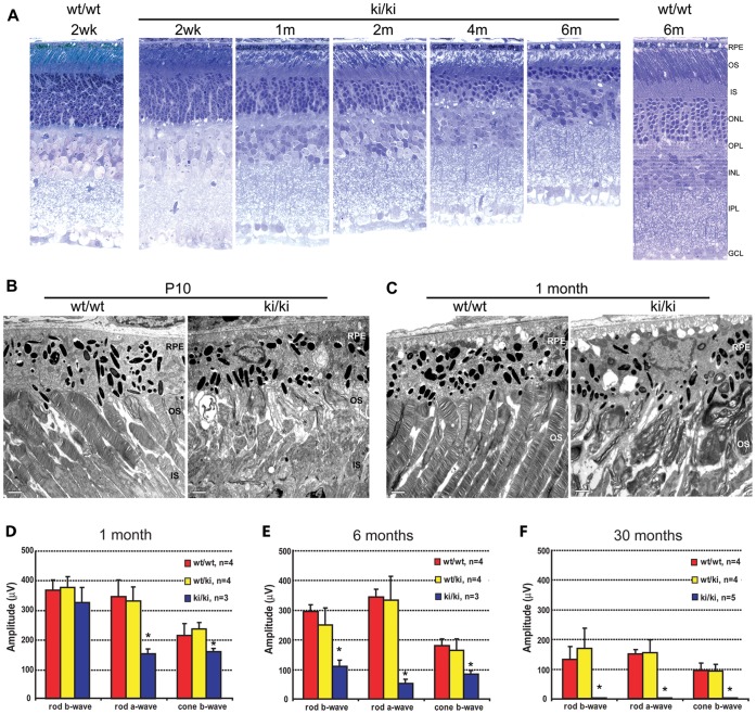
Figure 3. Retinal degeneration phenotype in the Rp1-Q662X mice. A.
Light microscopic images of semi-thin sections of retinas from wild-type (wt/wt) and homozygous Rp1-Q662X knock-in mice (ki/ki) of the ages indicated. Note that thinning of the outer nuclear layer in the ki/ki mice is evident at 1 month, and progresses so that by 6 months of age only 2–3 rows of photoreceptor nuclei remain. Note also the disorganization and shortening of the photoreceptor outer segments in the ki/ki mice. (GCL, ganglion cell layer; INL, inner nuclear layer; IPL, inner plexiform layer; IS, inner segment; ONL, outer nuclear layer; OPL, outer plexiform layer; OS, outer segment; RPE, retinal pigment epithelium; 400× magnification for all images). B, C. Electron micrographs of retinas from wt/wt and ki/ki mice at 10 days (B) and 1 month of age (C). At 10 days of age small packets of enlarged disc membranes parallel to the axis of the axonemes replaced the normal perpendicularly orientated stacks of discs observed in control animals. These defects of outer segments were even more evident at 1 month of age, with disorganized membranous whirls in place of outer segments, compared to the well aligned, uniform discs in the control retina (IS, inner segments; OS, outer segments; RPE, retinal pigment epithelium; bars = 2 µm). D–F. Average rod and cone ERG amplitudes for wild-type (wt/wt), heterozygous (wt/ki) and homozygous (ki/ki) Rp1-Q662X knock-in mice. The amplitudes + SD are shown for mice of the three genotypes at 1 month (D), 6 months (E) and 30 months of age (F). Significant differences (P<0.05) indicated by *. Note that the rod-a waves of the ki/ki mice were significantly decreased at 1 month of age. By 6 months of age, all measures of rod and cone function were decreased in the ki/ki mice. By 30 months of age, no recordable ERG responses were detected.

