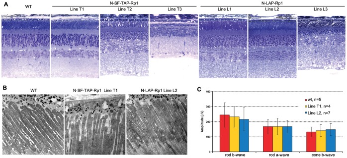
Figure 5. Retinal phenotypes of N-SF-TAP-Rp1 and N-LAP-Rp1 transgenic mice.
A. Retinal histology from 1-year-old mice of the genotypes indicated. Note that the retinal structure of the N-SF-TAP-Rp1 line T1 and N-LAP-Rp1 lines L1 and L2 is normal. In contrast, there is photoreceptor degeneration evident in N-SF-TAP-Rp1 lines T2 and T3, and N-LAP-Rp1 line L3, with loss of photoreceptor nuclei and shortening of photoreceptor outer segments. (INL, inner nuclear layer; IS, inner segment; ONL, outer nuclear layer; OS, outer segment; 400× magnification for all images). B. Ultrastructure of photoreceptor sensory cilia in one-year old N-SF-TAP-Rp1 line T1 and N-LAP-Rp1 line L2 mice, compared to that of wild-type littermate control. Note that the structure of the PSC and organization of the outer segment discs are normal in both transgenic lines, consistent with the histology shown in A. C. Amplitudes of ERG responses from 1-year-old N-SF-TAP-Rp1 line T1 and N-LAP-Rp1 line L2 mice, compared to that of wild-type littermate controls. Note that the rod and cone ERG amplitudes are normal in both lines of transgenic mice.

