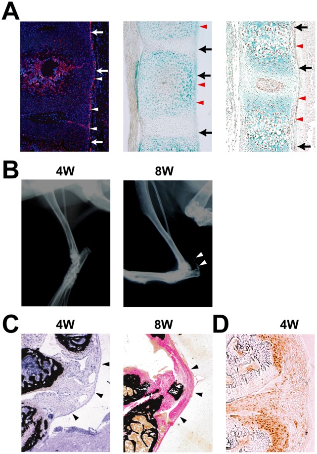
Figure 1. Expression of Runx2 in calcified ligament.
A, Runx2 expression (arrowheads) in the posterior longitudinal ligament (arrows). In situ hybridization of Runx2 in wild-type (WT) mouse vertebrae at birth (left). LacZ staining in WT mouse vertebrae at birth (middle). Immunohistochemistry of Runx2 in WT mouse vertebrae at embryonic day 16.5 (right). B, Radiographic assessment of the development of calcification of the ligament in an Enpp1ttw/ttw mouse at 4 and 8 weeks of age. Note an appearance of calcification at 8weeks (arrowheads) C, Histological assessment of the cruciform ligament (arrowheads) at the atlanto-occipital area in an Enpp1ttw/ttw mouse at 4 and 8 weeks of age. D, Immunohistochemical staining of Runx2 at the posterior longitudinal ligament in an Enpp1ttw/ttw mouse at 4 weeks of age. Note that Runx2 was expressed in an area corresponding to the prospective calcification.

