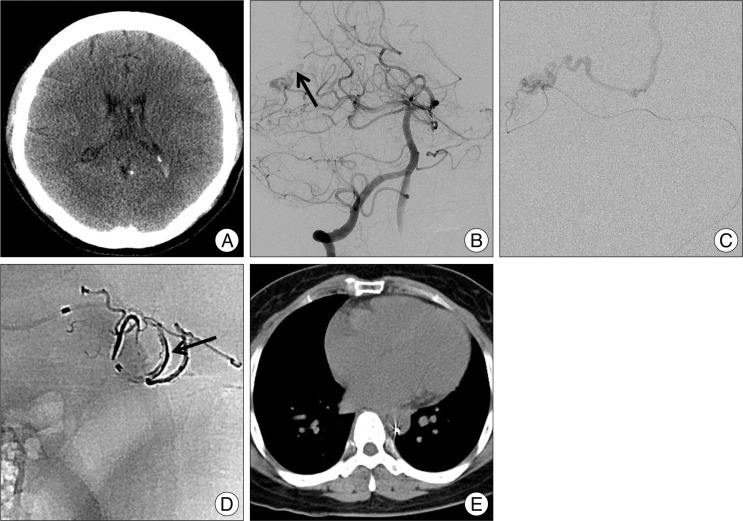
Fig. 1.
A : Non contrast enhanced brain computed tomography shows small amount of intraventricular hemorrhage in both lateral ventricle. B : Right vertebral artery angiography reveals small arteriovenous malformation (AVM) fed by right superior cerebellar artery braches and draining through internal cerebral vein (arrow). C : Superselective angiography demonstrates angioarchitecture of the AVM. D : Postprocedural radiography shows Onyx material. Black arrow indicates the cast of Onyx. E : Abdomino-pelvic computed tomography 2 years after embolization shows peripheral wall reposition of the retained microcatheter in descending aorta.

