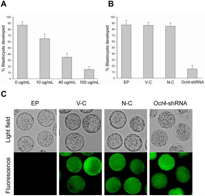
Figure 6. Electroporation of Oct4-specific shRNA expression vectors.
(A) Blastocyst development following electroporation of zygotes with custom Oct4-specific shRNA expression vectors at different concentrations and cultured for 3.5 d. (B) Blastocyst development following electroporation of zygotes with different shRNA expression vectors with a concentration of 100 µg/ml. (C) Morphological appearance of electroporated zygotes obtained from the control and Oct4-shRNA groups after being cultured for 3.5 d. Morphology (top) and fluorescence (bottom). The original magnification was ×100.

