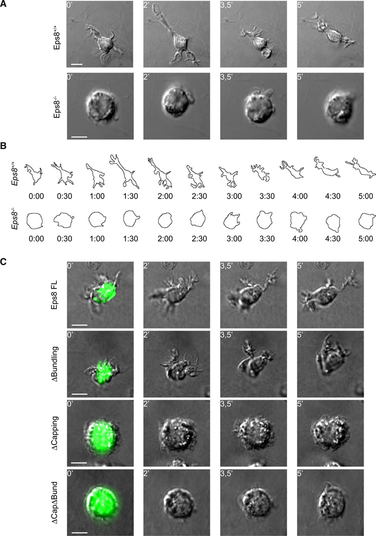
Figure 6. Eps8 Capping Activity Is Required for the Extension of Elongated and Persistent Cell Protrusions of BMDCs in 3D.
(A and B) Eps8−/− BMDCs fail to extend elongated cell protrusions and to migrate in 3D upon CCL19 stimulation.
(A) LPS-treated WT (Eps8+/+) and Eps8−/− (Eps8−/−) BMDCs were embedded in 1.6 mg/ml collagen type I chamber slides. CCL19 (1.2 µg/ml) was added to the top of the polymerized matrix before sealing the chamber. BMDC motility was recorded by spinning disk confocal microscopy. Still images from Movie S4 are shown. Scale bars represent 15 µm or 10 µm.
(B) Morphology outlines of WT (Eps8+/+) and Eps8−/− (Eps8−/−) BMDCs were obtained by morphometric analysis of 3D maximal projections. A time sequence of representative migratory WT (Eps8+/+) and Eps8−/− (Eps8−/−) BMDCs is shown.
(C) Eps8 capping, but not bundling activity, is required to restore Eps8−/− BMDC protrusions in 3D. Eps8−/− (Eps8−/−) BMDCs were transfected with either full-length (FL) GFP-Eps8 or the indicated mutants and treated with LPS and embedded in 1.6 mg/ml collagen type I matrix. CCL19 (1.2 µg/ml) was added in the upper part of the polymerized gel. BMDC protrusive activity was recorded as described above (see also Movie S5). Still images from representative movies are shown. At least 30 cells/experiment/condition of three independent BMDC preparations were analyzed by time lapse. The scale bar represents 10 µm.

