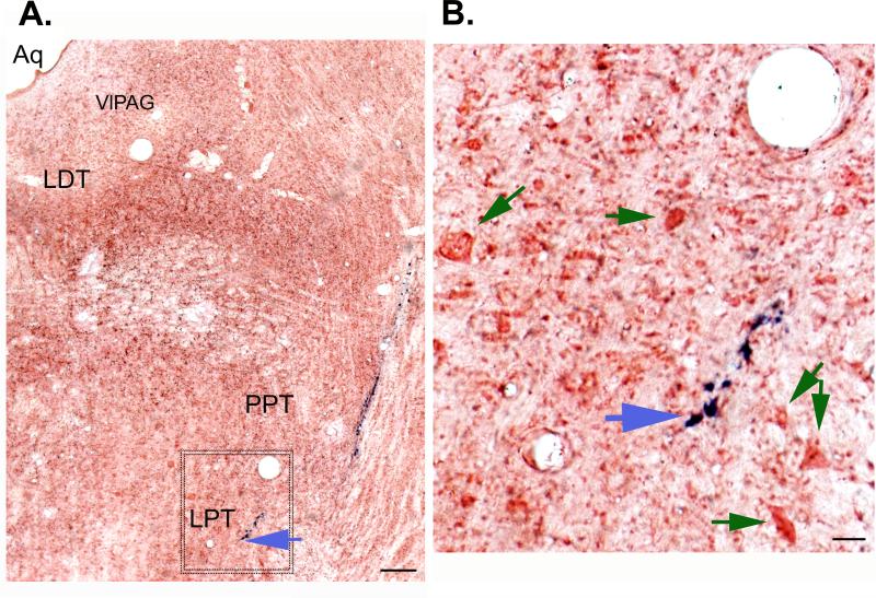
Figure 4.
A. Histological image showing the track of the recording electrode targeted to the lateral pontine tegmentum (LPT) where a phasic active wake (AW) spinally-projecting neuron was recorded. The blue mark (ferrous deposits) track (blue arrow) is from the recording electrode and is the result of the Pearl's Prussian-blue reaction seen against the background of the neutral red stain. B. Image shows an enlarged view of the area shown (box) in the figure 2A. Recording were done from the cells (green arrows) close to the electrode track. Bar in A. 50μm; B. 10 μm.

