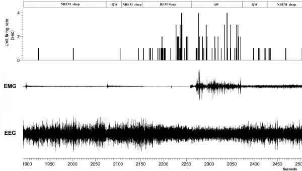
Figure 8. An example of a tonic firing AW and REM active neuron.
The neuron exhibits a higher firing rate irrespective of motor behavior in AW (higher EMG amplitude) or the REM sleep state (loss of motor activity) compared to QW and NREM sleep. This neuron was within the PPT region as verified by the electrode track.

