Figure 1.
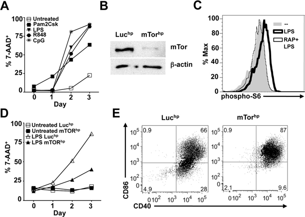
Inhibition of mTOR expression prolongs DC lifespan and promotes expression of costimulatory molecules CD40 and CD86. (A) DCs were pulsed with Pam2CSK4, LPS, R848, or CpG for 6 hours and then washed and cultured in complete medium. Cell viability was monitored daily by FACS analysis of 7-AAD staining of CD11c+ cells. (B) Western blot for mTOR and β-actin protein in Luciferase hpRNA (Luchp) or mTOR hpRNA (mTORhp) -transduced DCs. (C) DCs were stimulated with media alone, LPS, or rapamycin + LPS for 30 minutes. Cells were subsequently fixed and stained for phosphorylated S6 protein as a molecular readout for mTOR activation and analyzed by FACS. (D) Luchp and mTORhp-transduced DCs were left untreated or stimulated with LPS and monitored daily for cell viability by analysis of 7-AAD staining of CD11c+ cells. (E) Luchp or mTORhp DCs were cultured with or without LPS for 24 hours and analyzed by FACS for CD40 and CD86 expression. All graphs in this figure represent mean values of replicate wells; all experiments were performed at least twice with similar results.

