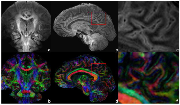
Figure 5.
Diffusion-weighted image of a coronal and sagittal slices (a,c) with b = 700 s/mm2 and corresponding FA DEC maps (b,d). Acquisition voxel size is 0.7/0.72/3.0 mm with TE of 70 ms, eight shots and five averages. An ROI is drawn over the mixed region of gray and white matter as a red box in (c) and (d). The ROI is shown enlarged in (e) and (f). Total scan time is 19 min for each of coronal and sagittal acquisitions.

