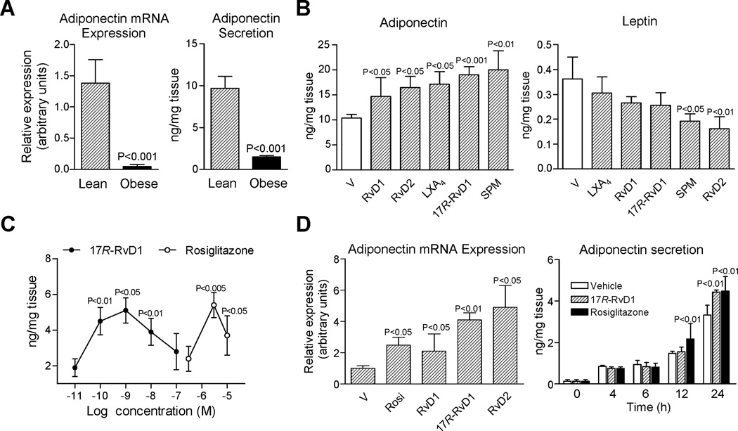
Figure 5. RvD1 and RvD2 potently induce adiponectin expression and secretion.
(A) mRNA expression and tissue levels of adiponectin in fat from lean and obese mice. (B) Adipose tissue explants were incubated ex vivo with vehicle (0.01% EtOH) or 10 nM RvD1, RvD2, LXA4 or 17R-RvD1 and a mixture of SPM (see text for details; 12 h at 37°C). Adiponectin and leptin levels in supernatants were quantitated by EIA. (C) Concentration-response curves for 17R-RvD1 (0.01–100 nM) and rosiglitazone (0.3–10 µM) on adiponectin secretion. (D) Changes in adiponectin expression in adipose tissue after 6 h of treatment with rosiglitazone (rosi, 3 µM) and equiconcentrations (10 nM) of RvD1, 17R-RvD1 and RvD2 (left panel); and time-response for 17R-RvD1 (10 nM) and rosiglitazone (3 µM) (right panel). Results are the mean±SEM of 5 separate experiments.

