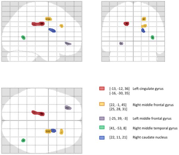
Figure 1.

Glass brain projections showing areas of significant (p<0.001; cluster size kE>50 voxels) gray matter volume reduction associated with exposure to alcohol in utero (exposed vs. controls) identified using optimized VBM. Gray matter regions were identified by converting MNI coordinates to Talairach space and labeled using Talairach Daemon searching for a gray matter label nearest the most significant voxel in the cluster.
