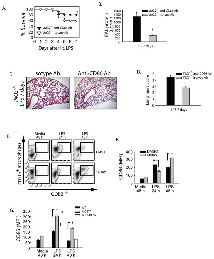
Fig. 9. iNOS regulates CD86 expression to regulate lung inflammation.
iNOS−/− mice were challenged with i.t. LPS and 48 hours after injury received either isotype Ab (Rat IgG) or anti-mouse CD86 (clone GL-1) on days +2, +4 and +6 after ALI. Survival (A) and BAL protein were evaluated in iNOS−/− mice after i.t. LPS. (C) Histological sections were stained with H & E (Original magnifications x20). (D) Histopathological mean lung injury scores from x20 sections (n=4–6 animals per group per group). The alveolar macrophage cell line (MH-S) was challenged with media or LPS (100 ng/ml) in the presence of DMSO or specific iNOS inhibitor 1400W (25 mM) at designated intervals. (E) Flow cytometry density plots show the relative expression of surface CD86 at designated intervals after LPS. (F) Mean fluorescence intensity (MFI) for CD86 expression was measured in MH-S cells and resident peritoneal macrophages (G) and compared between groups at intervals. Values expressed as mean ± SEM; † *, p<0.05 log-rank test (mortality curves) and unpaired Student’s t test (for other injury parameters).

