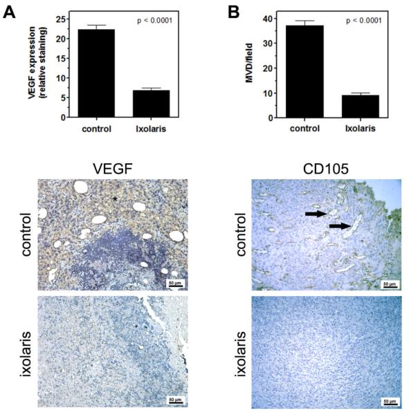
Figure 3. Ixolaris treatment reduces angiogenesis in primary melanoma tumors.
(A) Quantification of VEGF staining in tumor masses was performed as described in the Methods section. Bars show the decrease of relative VEGF staining in the primary tumors of animals treated with ixolaris (n=5; 6.8 ± 3.3) as compared to the control group (n=5; 22.4 ± 5.4). Representative images of VEGF staining (asterisk) are shown below. (B) Vessel density was evaluated in CD105-stained sections of the primary tumors, as described in the Methods section. Bars show the decrease of CD105 expression in the primary tumors of animals treated with ixolaris (n=5; 9.1 ± 3.5) as compared to the control group (n=5; 37.2 ± 6.1). Representative images of CD105 staining (arrows) are shown below. Values are given as the mean ± SD of each group.

