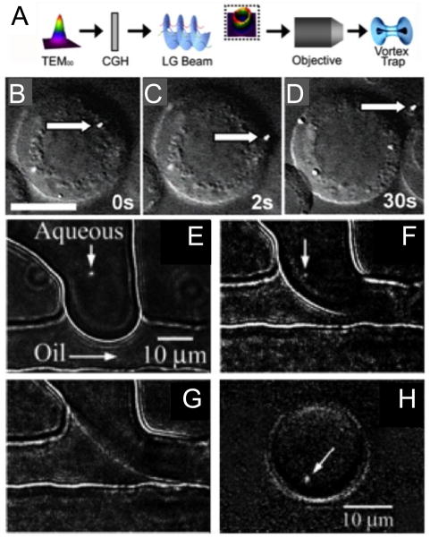
Figure 3.
A) Schematic showing the conversion of a Gaussian beam (TEM00) into that of an optical vortex beam (Laguerre-Gaussian (LG) beam) by using a computer generated hologram (CGH) and B–D) a sequence of images showing the removal of a fluorescent lysosome (stained with Lysotracker Green dye) from a B-lymphocyte. Scale bar = 10 μm in panel B. Reproduced with permission from Ref. [34]. E–H) Sequence of images showing the droplet encapsulation of a single mitochondrion. We visualized under fluorescence and optically manipulated a single mitochondrion stained with Mitotracker Green FM at the interface of the two fluids (E, F). Upon application of a pressure pulse to the microchannels (F, G), the mitochondrion was carried away by the flow as the droplet was sheared off (H). Reproduced with permission from Ref. [35].

