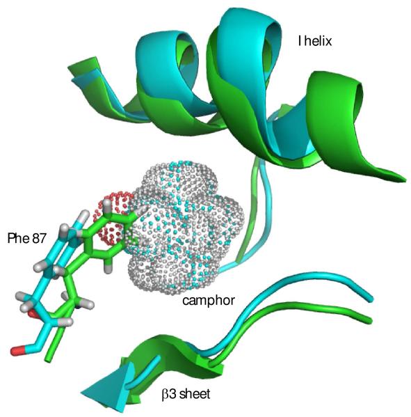
Figure 6.
Contraction of active site in REP1 (green) from best-fit backbone alignment to 2L8M (cyan). Camphor from 2L8M is shown as a van der Waals dot surface. Camphor-bound 2L8M structure is shown in cyan. Note that the side chain of Phe 87 partially occupies volume vacated by camphor. The side chains of Leu 244 and Val 247 also partially occlude the space occupied by camphor (see text). Unless otherwise noted, structural figures are generated using PyMOL (35).

