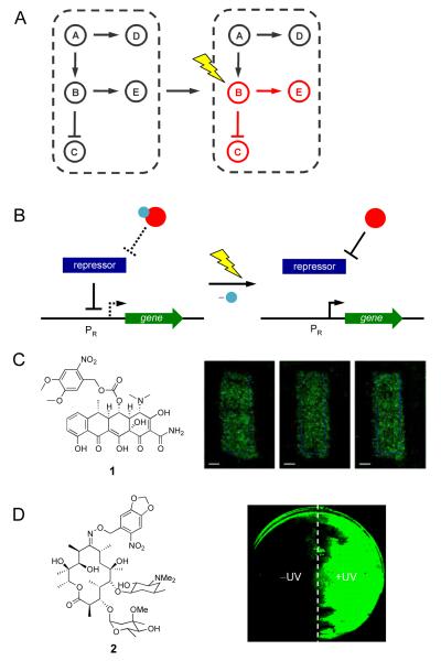
Figure 1.
(A) Light enables precise spatial and temporal activation of genetic circuits, even the activation of specific nodes in a natural or synthetic network. (B) Control over gene expression with photocaged small molecule inducers of transcription. The photocaging group is represented by a blue sphere and the small molecule inducer is represented by a red sphere. When the small molecule is caged, the repressor protein will bind the promoter PR. After UV irradiation the caging group is removed and the small molecule will bind the repressor, which releases PR, and allows for transcription to occur. (C) NvOC-Dox (1) was used to create photolithographic images onto an NIH 3T3 monolayer expressing GFP. (D) Photocaged erythromycin (2) was used in the spatial control of a light activated logic gate; cells treated with 2 where one half of the plate was exposed to UV light. Adapted with permission from the American Chemical Society, © 2010, from [17] and the Royal Society of Chemistry, © 2011, from [18].

