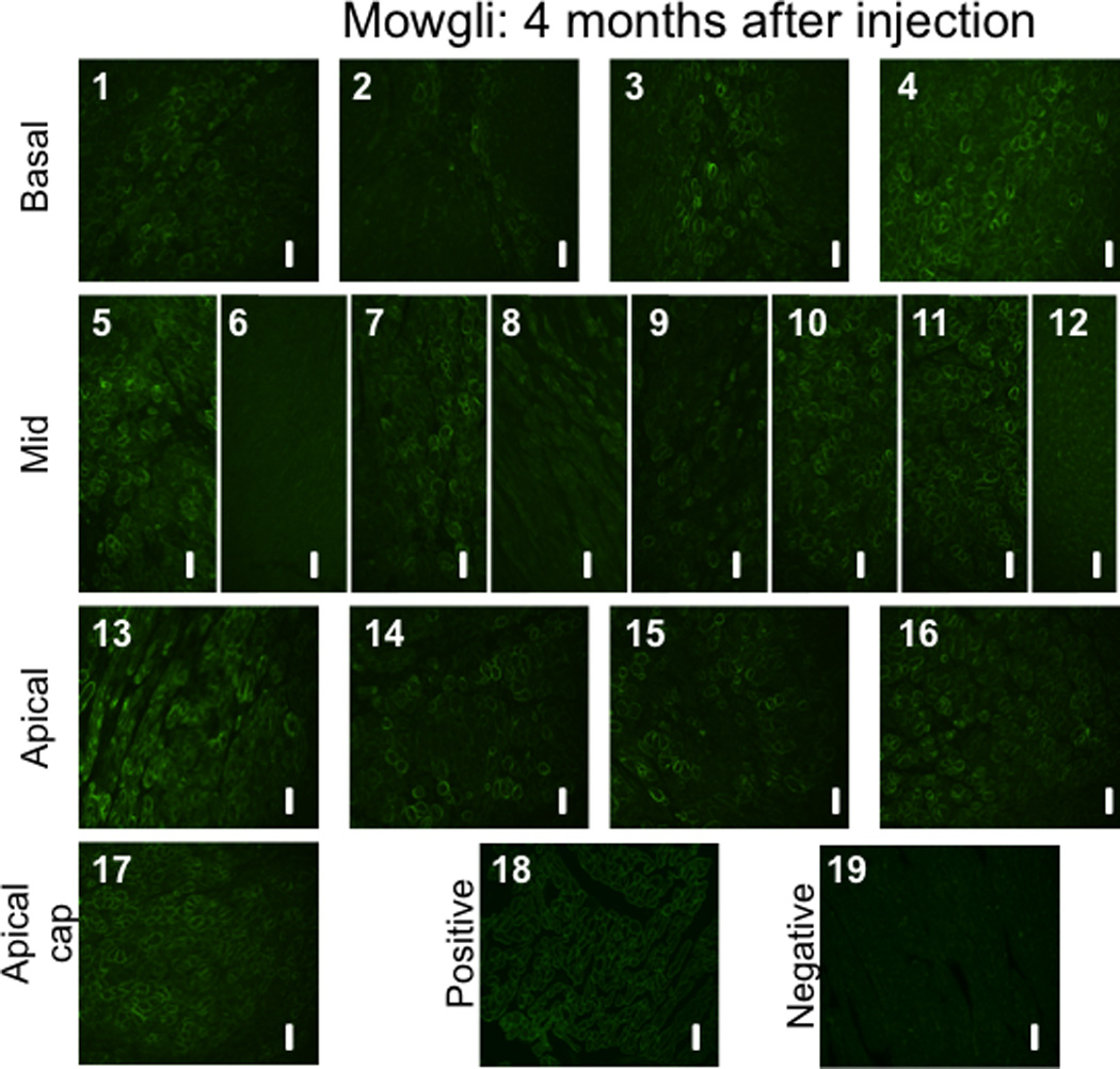
Figure 10. Immunohistochemical staining to dystrophin proteins in GRMD canines treated with rAAV-U7smOPT.

(1–17) Slides 1 to 17 (excluding slide 6) demonstrate various levels of positive staining for dystrophin in samples collected randomly from each of the 17 pre-defined left ventricular myocardial segments. The positive staining is typically located at the cell membrane, corresponding to the location of the functional dystrophin protein. As controls, tissues from (18) normal dog (positive control) and (19) untreated GRMD dog (negative control) were used. All images were acquired at a 20X magnification. The scale bars represents 50 µm.
