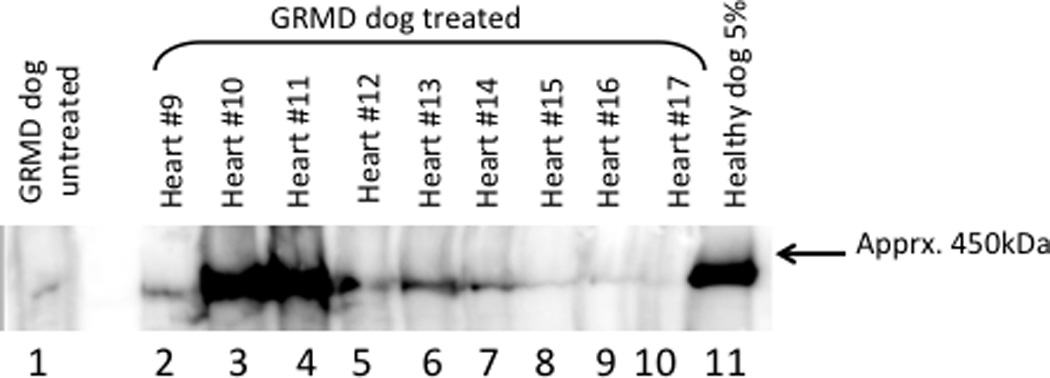
Figure 9. Western blot analysis of heart protein.

Soluble protein extracts prepared from randomly selected 20µm thick frozen sections were fractionated by SDS-polyacrylamide gel electrophoresis and transferred to PVDF membrane. The heart segments are indicated and the positive control consists of 5% normal dog heart protein extract in untreated GRMD heart protein extract. The negative control (lane 1) is untreated GRMD heart protein. The low level dystrophin protein signal was observed reproducibly and is likely the result of the so-called “revertant” myocytes in the GRMD dogs. Presumably, aberrant splicing restores the dystrophin open reading frame in a relatively small percentage of cells resulting in a natural form of exon skipping.
