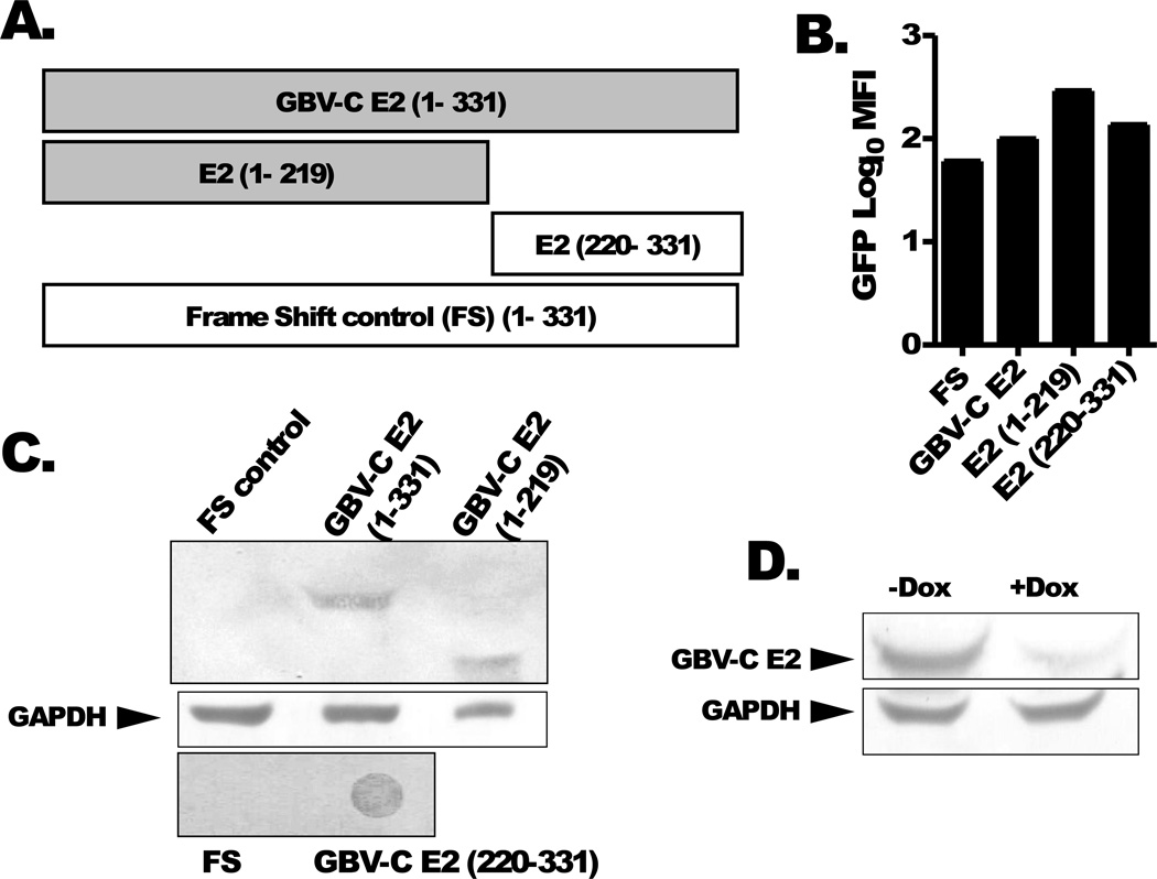
Figure 1. GBV-C E2 proteins and controls.
Schematic of GBV-C envelope protein E2 (nt 1167–2161; 331aa), N-terminal deletion mutant (aa 1–219; nt 1167–1824) and C terminal deletion mutant (aa 220–331; nt 1824–2161) and the 1–331 sequence with a frame shift inserted to abolish E2 expression (FS control). All of the recombinant proteins contained a C-terminal poly-histidine tag (A). Cell lines expressing the shaded constructs inhibited IL-2 release following T cell receptor activation, while non-shaded constructs did not. GFP expression was measured in the various cell lines (B). Immunoblot analysis using anti-His antibodies demonstrated recombinant GBV-C E2 protein expression in cell lines and deletion mutants (E2 1–219; E2 220–331) (panel C). The frameshift (FS) negative control is shown. GBV-C E2 protein expression was reduced more than 80% by maintaining Jurkat cells in doxycycline for 5 days (dox, 1 µg/mL) (D).

