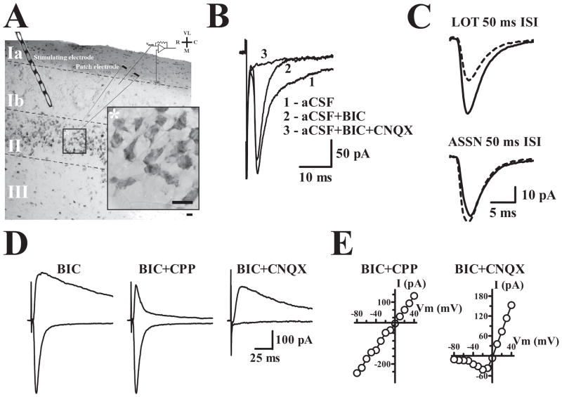
Figure 1. Physiological and anatomical properties of principal cells in APC.
A. 30 μm thick APC section stained with Cresyl violet and resectioned from a 300 μm thick 3–6 months old WT slice used in physiological recordings. ASSN synapses were stimulated with an electrode placed in layer Ib. Calibration bar, whole section: 100μm; inset: 10 μm. VL, ventrolateral; R, rostral; C, caudal; M, medial. B. Evoked recordings were initially obtained at −80 mV. Bicuculline (BIC) was included in the artificial cerebrospinal fluid (aCSF) perfusate to block inhibitory currents and reveal AMPA receptor mediated currents sensitive to AMPA receptor antagonist CNQX. C. Paired-pulse stimulation produces robust facilitation of lateral olfactory tract synapses in layer Ia and a small depression at ASSN synapses in layer Ib. Dashed line traces represent the initial evoked response and solid line traces represent the event elicited by the second pulse presented at an IPI of 50 ms. D. Evoked currents at −80 mV and +40 mV illustrate the pronounced voltage-dependent (NMDA) current component at positive potentials. AMPA and NMDA components of evoked currents were isolated by CPP and CNQX respectively. E. Current-Voltage plots for cells in which the AMPA (BIC+CPP) or NMDA (BIC+CNQX) currents were isolated using the indicated drugs.

