
Fig. 6.
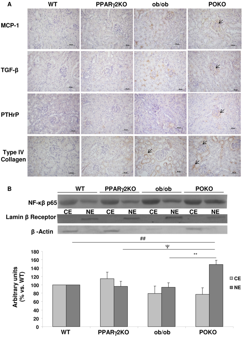
Inflammation markers and renal injury in the four genotypes. (A) Immunostaining for MCP-1, TGFβ, PTHrP and type IV collagen in the kidney from male WT, PPARγ2 KO, ob/ob and POKO mice at 4 weeks (n=4–5). Original magnification: 400×; scale bars: 50 μm. (B) Representative immunoblot for cytosolic (CE) and nuclear (NE) p65 NFκB protein in renal extracts from WT, PPARγ2 KO, ob/ob and POKO 4-week-old male mice. Levels were normalized to β-actin in the cytosolic fraction and lamin-β receptor in the nuclear fraction. Each value is the relative optical intensity of each band normalized as a percentage of that of the WT group. Values are represented in graphic (n=5–7). Data are means±s.e.m. **P<0.01 POKO vs ob/ob; ##P<0.01 POKO vs WT; ΨP<0.05 POKO vs PPARγ2 KO.