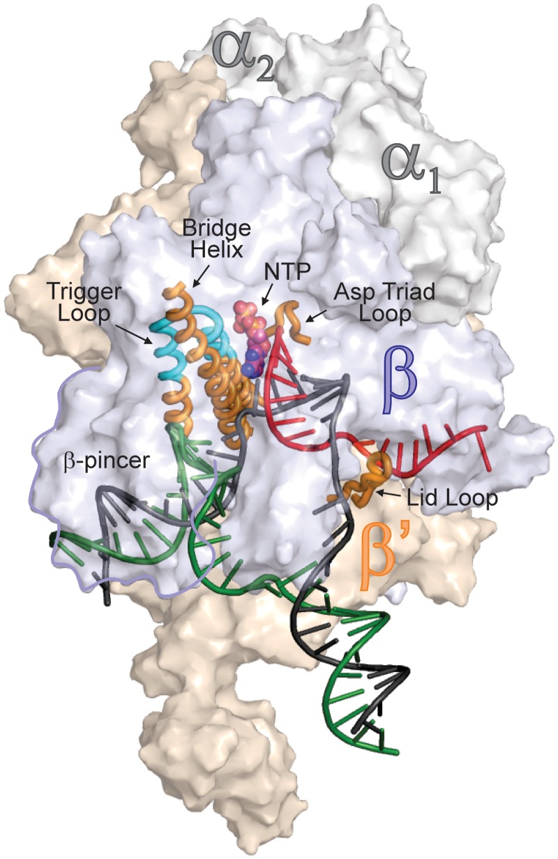
Figure 1.
An overview of bacterial TEC structure. β (light blue), β′ (wheat) and two (gray) subunits are depicted as transparent surfaces; the non-essential ω subunit is obstructed by β and β′ subunits. Template DNA, non-template DNA and RNA are depicted in black, green and red, respectively. The β′ subunit elements discussed throughout the manuscript are depicted as cartoons: the folded TL is colored cyan; the bridge helix (BH), lid loop, TL base and catalytic aspartate triad loop are colored orange. The β-pincer domain is outlined. Substrate NTP atoms are shown as spheres. RNAP is drawn using coordinates of Thermus thermophilus TEC PDB ID 2O5J, and nucleic acids are taken from the E. coli TEC model (15).

