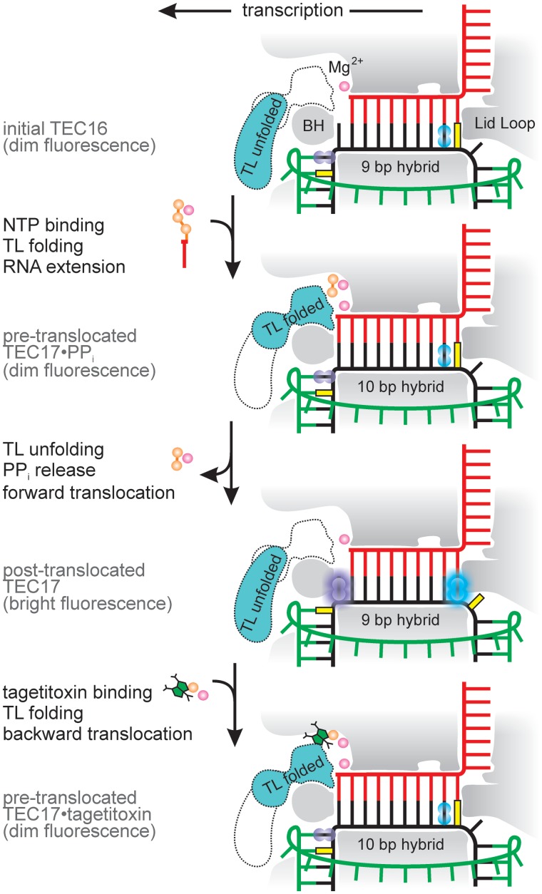
Figure 2.
Overview of the experimental setup used in this study. RNAP is shaded gray. Two alternative conformations of the TL domain are indicated, with the predominant conformation shaded in turquoise (see ‘Discussion’ section). Template DNA, non-template DNA and RNA are depicted in black, green and red, respectively. Although presented as a bifluorophore for compact representation, each TEC in our experiments possessed only one of the indicated fluorophores: either guanine analog 6-MI (cyan) at i − 7 or adenine analog 2-AP (violet) at i + 2 (for the initial TECs). Guanine, serving as a quencher, is depicted in yellow.

