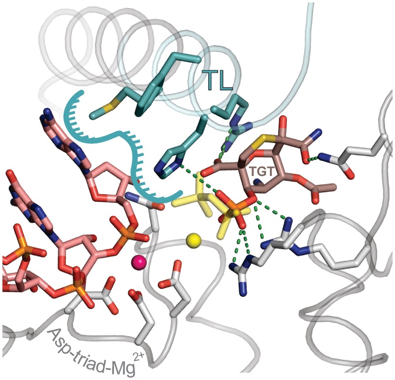
Figure 8.
Structural model of the closed TEC–TGT complex. Phosphate and carboxylate groups of TGT (brown) overlap with the PPi molecule (yellow). TL is colored turquoise and the rest of RNAP is colored gray. RNA is colored rose. The surface contributed by TL residues for interaction with RNA 3′ NMP is contoured by a turquoise line. Asp triad residues and selected side chains that interact with TGT and the 3′ RNA nucleobase are shown as sticks. Active site Mg2+ ions coordinated by the Asp triad and PPi are depicted as magenta and yellow spheres, respectively. TGT–protein contacts are indicated as green dashed lines.

