


Abstract
Osteoporosis is considered a serious debilitating disease. Cosmos caudatus (ulam raja), a plant containing antioxidant compounds and minerals, may be used to treat and prevent osteoporosis. This study determines the effectiveness of C. caudatus as bone protective agent in postmenopausal osteoporosis rat model. Thirty-two female rats, aged 3 months old, were divided into 4 groups. Group one was sham operated (sham) while group two was ovariectomized. These two groups were given ionized water by forced feeding. Groups three and four were ovariectomized and given calcium 1% ad libitum and force-fed with C. caudatus at the dose of 500 mg/kg, respectively. Treatments were given six days per week for a period of eight weeks. Body weight was monitored every week and structural bone histomorphometry analyses of the femur bones were performed. Ovariectomy decreased trabecular bone volume (BV/TV), decreased trabecular number (Tb.N), and increased trabecular separation (Tb.Sp). Both calcium 1% and 500 mg/kg C. caudatus reversed the above structural bone histomorphometric parameters to normal level. C. caudatus shows better effect compared to calcium 1% on trabecular number (Tb.N) and trabecular separation (Tb.Sp). Therefore, Cosmos caudatus 500 mg/kg has the potential to act as the therapeutic agent to restore bone damage in postmenopausal women.
1. Introduction
Estrogen deficiency increases the risk of developing osteoporosis. Estrogen was found to have antioxidant properties [1] and was also shown to increase the expression of glutathione peroxidase in osteoclasts [2], an enzyme which is responsible for the degradation of hydrogen peroxide. Estrogen deficiency will reduce the expression of the enzyme and renders the bone susceptible to hydrogen peroxide attacks.
In osteoporosis, lipid peroxidation is increased due to the reduction in antioxidants [3], and reactive oxygen species are found to play a role in bone metabolism [4]. Free radicals have also been shown to be cytotoxic to osteoblastic cells [5]. Loss of estrogens accelerates the effects of aging on bone by decreasing defence against oxidative stress which leads to bone loss [6].
Since free radicals and lipid peroxidation are involved in bone metabolism and may be the culprit in causing bone loss, substances having antioxidative activities can overcome the detrimental effects. Our previous studies have shown the beneficial effects of palm-oil derived tocotrienols in several experimental osteoporosis, ovariectomized [7], steroid induced [8], and nicotine induced [9]. The effects of palm-oil derived tocotrienols may be attributed to its antioxidative activities.
Thus, in finding alternatives in the treatment of osteoporosis, a local plant, Cosmos caudatus or locally known as “ulam raja” (King's salad), is of consideration. Previous study has shown that this plant has antioxidative activities [10]. It contains phenolic compounds that contribute to the color, antioxidant, and anticarcinogenic properties of the plants. For every 100 g of Cosmos caudatus, the total phenolic compound is 21.41 mg. It is also found that Cosmos caudatus had extremely high antioxidant capacity of about 2,400 mg l ascorbic acid equivalent antioxidant capacity (AEAC) per 100 g of fresh sample [11]. It is also believed that Cosmos caudatus promote the formation of healthy bones [12]. Thus, we hypothesized that Cosmos caudatus may exert protective effects on bone of ovariectomized rats which is a suitable animal model for studying postmenopausal osteoporosis. In this study, the effects of Cosmos caudatus on structural bone histomorphometry were determined.
2. Materials and Methods
2.1. Animals and Treatment
Thirty-two young adult (3 months) female Wistar rats, weighing 190 g–260 g, were obtained from the Laboratory Animal Resource Unit, Faculty of Medicine, Universiti Kebangsaan Malaysia. Rats were randomly assigned to four groups with eight rats in each group. Group 1 was sham operated (sham) while the second was ovariectomized-control group (OVX). The third and fourth groups were ovariectomized and treated with calcium 1% (Ca) ad libitum and force-fed with 500 mg/kg C. caudatus extract (CC), respectively. Treatment was given six days a week for eight weeks and body weight was recorded weekly. The study was approved by the Universiti Kebangsaan Malaysia Animal Ethics Committee with the approval code of PP/FAR/2008/NORAZLINA/12-AUGUST/225-SEPT-2008-AUG-2009.
2.1.1. Diet, Cosmos caudatus, and Calcium 1%
All rats received normal rat chow obtained from Gold Coin, Malaysia. The composition of rat chow is shown in Table 1. The aqueous extract of C. caudatus with the concentration of 500 g/300 mL was prepared by School of Chemical Sciences & Food Technology, Faculty of Science and Technology, Universiti Kebangsaan Malaysia using water extraction method which was previously described [10]. The 500 mg/kg dose was prepared by mixing C. caudatus with deionized water in ratio 3 : 7. Calcium 1% solution was prepared by mixing 1 g of hemicalcium lactic acid (Sigma Chemical CO., USA) with 100 mL deionized water.
Table 1.
Composition of rat chow (Golden Coin, Malaysia).
| Composition | Amount/percentage |
|---|---|
| Crude protein | 21–23% |
| Crude fibre (max) | 5.0% |
| Crude fat (min) | 3.0% |
| Moisture (max) | 3.0% |
| Calcium | 0.8–1.2% |
| Phosphorus | 0.6–1.0% |
| Nitrogen free extract | 49.0% |
| Vitamin A | 10 M.I.U. |
| Vitamin D3 | 2.5 M.I.U |
| Vitamin E | 15 g |
| Vitamin K | trace |
| Vitamin B12 | trace |
| Thiamine | trace |
| Riboflavin | trace |
| Pantothenic acid | trace |
| Niacin | trace |
| Pyridoxine | trace |
| Choline | trace |
| Santoquin | trace |
| Microminerals | trace |
2.2. Ovariectomy
Before the surgery, the rats were anesthetized with Ketamil and Ilium Xylazil-20 (Troy Laboratories PTY, Australia), given intraperitoneally, at 1 : 1 ratio. A vertical incision was made approximately 15 cm in the abdomen using a sterilized sharp knife. The right and left ovaries were cut and removed. Before the ovaries were cut, the fallopian tubes were tied to prevent bleeding. The muscle layer under the skin was stitched up by sterile and soluble suture (Serafit, Germany). Then, the outer layer of skin was sewn with nonwater soluble suture (Seralon, Serag Wiessner, Germany). The procedure for sham operated rats was just the same with ovariectomized rats, but both of the ovaries were not removed. Rats were left recuperating for 1 week before commencing the treatment.
2.3. Bone Histomorphometry
Upon sacrifice, the distal part of the femur was fixed with 70% ethanol and undergoes undecalcified bone preparations. The bone samples were embedded in polymer methyl methacrylate according to Difford [13], sectioned at 9 μm thickness using a microtome, stained using von Kossa method [14], and analyzed using an image analyzer with the Video Test-Master software. The parameters measured were trabecular bone volume (BV/TV), trabecular thickness (Tb.Th), trabecular number (Tb.N), and trabecular separation (Tb.Sp). All measurements were performed randomly at the metaphyseal region, which was located 3–7 mm from the lowest point of the growth plate and 1 mm from the lateral cortex [15]. The selected area is the secondary spongiosa area, which is rich in trabecular bone. All parameters were measured according to the American Society of Bone Mineral Research Histomorphometry Nomenclature Committee [16].
2.4. Statistical Analysis
Results were presented as mean ± standard error of the mean (SEM). All data were analysed using the Statistical Package for Social Sciences software. The Kolmogorov-Smirnov test was used for normality. ANOVA followed by Tukey's HSD tests were used for normally distributed data while Kruskal-Wallis and Mann-Whitney tests were used for not normally distributed data.
3. Results
Figure 1 shows the body weight of all groups throughout the study period. Ovariectomized group gained weight and was significantly different compared to sham-operated group beginning from week 3. Similar findings were observed in the calcium supplemented group. Group supplemented with C. caudatus also showed weight gain but did not differ compared to the sham-operated group.
Figure 1.

Body weight of rats for all treatment groups. *Indicates significant difference (P < 0.05) compared to SO group. SO, sham operated; Ovx, ovariectomized; CC, ovariectomized and supplemented with 500 mg/kg Cosmos caudatus; Ca, ovariectomized and supplemented with 1% calcium.
Figure 2 shows the photomicrographs of trabecular bone with von Kossa staining for the different treatment groups. Ovariectomy caused a reduction in bone volume (BV/TV) and an increase in trabecular separation (Tb.Sp) compared to the control group (Figures 3 and 4, resp.). Administration of C. caudatus was able to improve bone volume and trabecular separation in ovariectomized rats (Figures 3 and 4, resp.) while calcium administration was only able to improve bone volume (Figure 3). In addition, the group supplemented with C. caudatus showed a higher trabecular number compared to ovariectomized group (Figure 5). No significant differences were seen in trabecular thickness (Figure 6).
Figure 2.

Photomicrographs of trabecular bone. Undecalcified section (100 x magnification) shows trabecular bone (stained black) using von Kossa method. SO, sham operated; Ovx, ovariectomized; CC, ovariectomized and supplemented with 500 mg/kg Cosmos caudatus; Ca, ovariectomized and supplemented with 1% calcium.
Figure 3.

Effects of Cosmos caudatus supplementation on bone volume in ovariectomized rats. Groups which share the same alphabet indicate significant difference (P < 0.05). SO, sham operated; Ovx, ovariectomized; CC, ovariectomized and supplemented with 500 mg/kg Cosmos caudatus; Ca, ovariectomized and supplemented with 1% calcium.
Figure 4.
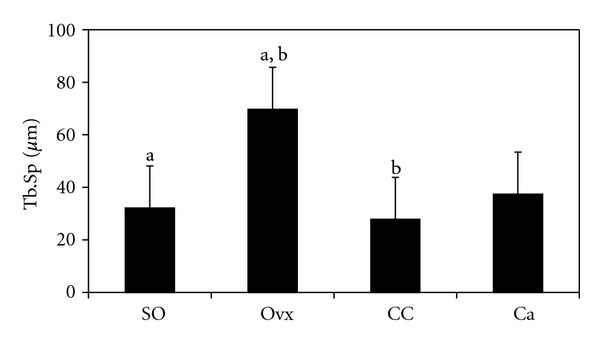
Effects of Cosmos caudatus supplementation on trabecular separation in ovariectomized rats. Groups which share the same alphabet indicate significant difference (P < 0.05). SO, sham operated; Ovx, ovariectomized; CC, ovariectomized and supplemented with 500 mg/kg Cosmos caudatus; Ca, ovariectomized and supplemented with 1% calcium.
Figure 5.
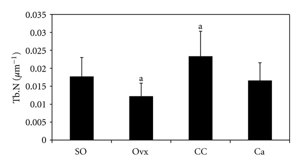
Effects of Cosmos caudatus supplementation on trabecular number in ovariectomized rats. Groups which share the same alphabet indicate significant difference (P < 0.05). SO, sham operated; Ovx, ovariectomized; CC, ovariectomized and supplemented with 500 mg/kg Cosmos caudatus; Ca, ovariectomized and supplemented with 1% calcium.
Figure 6.

Effects of Cosmos caudatus supplementation on trabecular thickness in ovariectomized rats. SO, sham operated; Ovx, ovariectomized; CC, ovariectomized and supplemented with 500 mg/kg Cosmos caudatus; Ca, ovariectomized and supplemented with 1% calcium.
4. Discussion
Ovariectomized rats have been used by researchers as the model for postmenopausal osteoporosis. Even though limitations exist, certain characteristics in the rat model mimic the bone changes in postmenopausal women and made the study of the human disease possible [17, 18]. Reduction in bone mineral density occurs two months after ovariectomy with greater loss seen in regions rich in trabecular bone [19].
Ovariectomized group showed increase in body weight which was statistically different compared to sham-operated group beginning from week 3 (Figure 1). This observation is common in estrogen-deficient animals since the deficient state induces hyperphagia in rats [20]. Group supplemented with calcium showed the same findings. However, rats given C. caudatus did not show a significant increase in body weight. Cosmos caudatus seemed able to prevent weight gain induced by ovariectomy. However, the exact mechanism of this occurrence is unknown.
In this study, ovariectomy caused loss of bone volume, increase in trabecular separation, and decrease in trabecular number (Figures 3, 4, and 5, resp.). The bone loss is reflected in the photomicrograph of the trabecular bone in which the ovariectomized group showed perforated and discontinued trabecular bone compared to the sham-operated group (Figures 2(a) and 2(b)). Similar findings were observed in other studies [21, 22]. However, trabecular thickness did not show any significant differences between the different groups. This is in contrast with other studies [23, 24] which used micro-CT in their studies as opposed to conventional histomorphometry in our study. In another study which used older rats, ovariectomy was also shown to cause a decrease in trabecular thickness [25]. Different method and different age range of the animals may contribute to the discrepancy seen in this study.
Estrogen deficiency is the major factor which affects bone in ovariectomized rats [26]. The deficiency state induces osteocytes apoptosis which further leads to increase in osteoclastic resorption [27]. Ovariectomy has been associated with increase in oxidative stress as evident by high malondialdehyde levels [28]. The condition of oxidative stress would eventually lead to bone loss [29].
Ovariectomized rats supplemented with C. caudatus showed improvements in bone volume, trabecular separation and trabecular number (Figures 3, 4, and 5, resp.). Photomicrograph of the trabecular bone of rats given C. caudatus appears similar to the sham-operated rats (Figures 2(a) and 2(c)). Cosmos caudatus is shown to contain flavonoids [30] and ascorbic acid [31]. It has also been shown to exert antioxidant activity [32]. This may contribute to the bone protective effects of C. caudatus observed in the present study.
The dose of C. caudatus in this study was chosen based on previous study which used C. caudatus at the dose of 100, 200, and 300 mg/kg. In the previous study, the effects of C. caudatus on bone biochemical markers and bone histomorphometry in ovariectomized rats were determined. It was found that C. caudatus at all doses was able to prevent the increase in interleukin-1 and pyridinoline as seen in the ovariectomized group. However, no significant changes were seen in the bone histomorphometry parameters (unpublished data). Thus, in the present study, a higher dose, 500 mg/kg, was used.
In an acute toxicity study done previously, a single dose of C. caudatus was given to male rats at the dose of 50, 500, and 2000 mg/kg. The rats were kept for 7 days before sacrifice. The higher dose of C. caudatus, that is, 2000 mg/kg, was found to increase liver enzymes but did not cause any changes on the haematological parameters such as clotting time, bleeding time, platelet levels, and white cell count (unpublished data). The observations above imply that C. caudatus at the dose of 500 mg/kg is not associated with side effects.
Calcium supplement in combination with vitamin D is recommended in the treatment regime for patients with osteoporosis [33] to reduce risk of nonvertebral fractures [34]. This combination therapy is considered essential but not sufficient for the treatment of osteoporosis and additional benefit may be obtained with the addition of antiresorptive or anabolic agent [35]. In the present study, calcium supplementation also reversed the effects of ovariectomy on bone volume (Figure 2) but failed to show significant changes in the other parameters. Similar findings were observed in previous study in which ovariectomized rats given calcium supplementation still had lower bone volume and trabecular number as compared to sham-operated rats [36]. In another study using ovariectomized rats, calcium supplementation is shown to improve bone fracture healing but failed to improve strength [37]. Another study showed that calcium supplementation suppresses bone formation in magnesium-deficient rats [38]. The findings above suggested that calcium may need to be present with other elements to exert beneficial effects on bone.
All rats received rat chow which contained 0.8–1.2% calcium. The Ca group received additional 1% calcium in drinking water. However, since the calcium was administered via drinking water, the amount of calcium ingested by the rats may vary between individual rats thus causing inconsistent results in the histomorphometry parameters mentioned above.
In the present study, we observed that C. caudatus improved bone structural histomorphometric parameters. In fact, C. caudatus seemed to yield better effects on bone in terms of trabecular separation and trabecular number (Figures 4 and 5). According to Nutriweb Malaysia [39], C. caudatus contains 270 mg calcium per 100 g of the plant. This composition, in addition to its antioxidative property, enables C. caudatus to have a greater effect in reversing bone changes induced by ovariectomy as opposed to calcium supplementation.
Further studies are required to establish the effects of Cosmos caudatus on bone such as the effects on dynamic histomorphometry, bone biomarkers, bone density, and bone calcium content. In addition, further studies are also required to ascertain the active compound and calcium content of the plant as well as its exact mechanism on bone.
5. Conclusion
Cosmos caudatus at the dose of 500 mg/kg reversed bone changes induced by ovariectomy. Thus, C. caudatus has the potential to be used as an alternative for the treatment of postmenopausal osteoporosis.
Acknowledgments
The authors would like to thank Mr. Muhamad Arizi Aziz for the technical support. The authors also expressed their gratitude to the Faculty of Medicine, Universiti Kebangsaan Malaysia for funding the research (Grant no. FF-193-2008).
References
- 1.Badeau M, Adlercreutz H, Kaihovaara P, Tikkanen MJ. Estrogen A-ring structure and antioxidative effect on lipoproteins. Journal of Steroid Biochemistry and Molecular Biology. 2005;96(3-4):271–278. doi: 10.1016/j.jsbmb.2005.04.034. [DOI] [PubMed] [Google Scholar]
- 2.Lean JM, Jagger CJ, Kirstein B, Fuller K, Chambers TJ. Hydrogen peroxide is essential for estrogen-deficiency bone loss and osteoclast formation. Endocrinology. 2005;146(2):728–735. doi: 10.1210/en.2004-1021. [DOI] [PubMed] [Google Scholar]
- 3.Maggio D, Barabani M, Pierandrei M, et al. Marked decrease in plasma antioxidants in aged osteoporotic women: results of a cross-sectional study. Journal of Clinical Endocrinology and Metabolism. 2003;88(4):1523–1527. doi: 10.1210/jc.2002-021496. [DOI] [PubMed] [Google Scholar]
- 4.Sontakke AN, Tare RS. A duality in the roles of reactive oxygen species with respect to bone metabolism. Clinica Chimica Acta. 2002;318(1-2):145–148. doi: 10.1016/s0009-8981(01)00766-5. [DOI] [PubMed] [Google Scholar]
- 5.Moreau MF, Chappard D, Lesourd M, Montheard JP, Basle MF. Free radicals and side products released during methylmethacrylate polimeryzation are cytotoxic for osteoblastic cells. Journal of Biomedical Materials Research. 1998;40:124–131. doi: 10.1002/(sici)1097-4636(199804)40:1<124::aid-jbm14>3.0.co;2-o. [DOI] [PubMed] [Google Scholar]
- 6.Almeida M, Han L, Martin-Millan M, et al. Skeletal involution by age-associated oxidative stress and its acceleration by loss of sex steroids. Journal of Biological Chemistry. 2007;282(37):27285–27297. doi: 10.1074/jbc.M702810200. [DOI] [PMC free article] [PubMed] [Google Scholar]
- 7.Norazlina M, Ima-Nirwana S, Gapor MT, Khalid BAK. Palm vitamin E is comparable to α-tocopherol in maintaining bone mineral density in ovariectomised female rats. Experimental and Clinical Endocrinology and Diabetes. 2000;108(4):305–310. doi: 10.1055/s-2000-7758. [DOI] [PubMed] [Google Scholar]
- 8.Ima-Nirwana S, Fakhrurazi H. Palm vitamin E protects bone against dexamethasone-induced osteoporosis in male rats. Medical Journal of Malaysia. 2002;57:133–141. [PubMed] [Google Scholar]
- 9.Norazlina M, Lee PL, Lukman HI, Nazrun AS, Ima-Nirwana S. Effects of vitamin E supplementation on bone metabolism in nicotine-treated rats. Singapore Medical Journal. 2007;48(3):195–199. [PubMed] [Google Scholar]
- 10.Huda-Faujan N, Noriham A, Norrakiah AS, Babji AS. Antioxidative activities of water extracts of some Malaysian herbs. ASEAN Food Journal. 2007;14(1):61–68. [Google Scholar]
- 11.Shui G, Leong LP, Shih PW. Rapid screening and characterisation of antioxidants of Cosmos caudatus using liquid chromatography coupled with mass spectrometry. Journal of Chromatography B. 2005;827(1):127–138. doi: 10.1016/j.jchromb.2005.07.029. [DOI] [PubMed] [Google Scholar]
- 12.Ismail S. Sayuran Tradisional Ulam dan Penyedap Rasa. Bangui, Central African Republic: UKM; 2000. [Google Scholar]
- 13.Difford J. A simplified method for the preparation of methyl methacrylate embedding medium for undecalcified bone. Medical Laboratory Technology. 1974;31(1):79–81. [PubMed] [Google Scholar]
- 14.Von Kossa J. Nachweis von Kalk. Beitrage zur pathologischen Anatomie und zur allgemeinen. Pathologie. 1901;29, article 163 [Google Scholar]
- 15.Baldock PAJ, Morris HA, Need AG, Moore RJ, Durbridge TC. Variation in the short-term changes in bone cell activity in three regions of the distal femur immediately following ovariectomy. Journal of Bone and Mineral Research. 1998;13(9):1451–1457. doi: 10.1359/jbmr.1998.13.9.1451. [DOI] [PubMed] [Google Scholar]
- 16.Parfitt AM, Drezner MK, Glorieux FH, et al. Bone histomorphometry: standardization of nomenclature, symbols, and units. Report of the ASBMR Histomorphometry Nomenclature Committee. Journal of Bone and Mineral Research. 1987;2(6):595–610. doi: 10.1002/jbmr.5650020617. [DOI] [PubMed] [Google Scholar]
- 17.Lelovas PP, Xanthos TT, Thorma SE, Lyritis GP, Dontas IA. The laboratory rat as an animal model for osteoporosis research. Comparative Medicine. 2008;58(5):424–430. [PMC free article] [PubMed] [Google Scholar]
- 18.Turner AS. Animal models of osteoporosis—necessity and limitations. European Cells and Materials. 2001;1:66–81. doi: 10.22203/ecm.v001a08. [DOI] [PubMed] [Google Scholar]
- 19.Ima-Nirwana S, Norazlina M, Khalid BAK. Pattern of bone mineral density in growing male and female rats after gonadectomy. Journal of ASEAN Federation of Endocrine Societies. 1998;16:21–36. [Google Scholar]
- 20.Eckel LA. The ovarian hormone estradiol plays a crucial role in the control of food intake in females. Physiology and Behavior. 2011;104(4):517–524. doi: 10.1016/j.physbeh.2011.04.014. [DOI] [PMC free article] [PubMed] [Google Scholar]
- 21.Guo HY, Jiang L, Ibrahim SA, et al. Orally administered lactoferrin preserves bone mass and microarchitecture in ovariectomized rats. Journal of Nutrition. 2009;139(5):958–964. doi: 10.3945/jn.108.100586. [DOI] [PubMed] [Google Scholar]
- 22.Washimi Y, Ito M, Morishima Y, et al. Effect of combined humanPTH(1-34) and calcitonin treatment in ovariectomized rats. Bone. 2007;41(5):786–793. doi: 10.1016/j.bone.2007.06.019. [DOI] [PubMed] [Google Scholar]
- 23.Zhang R, Liu ZG, Li C, et al. Du-Zhong (Eucommia ulmoides Oliv.) cortex extract prevent OVX-induced osteoporosis in rats. Bone. 2009;45(3):553–559. doi: 10.1016/j.bone.2008.08.127. [DOI] [PubMed] [Google Scholar]
- 24.Yao X, Chen H, Emura S, Otake N, Shoumura S. Effects of hPTH (1-34) and Gosha-jinki-gan on the trabecular bone microarchitecture in ovariectomized rat tibia. Okajimas Folia Anatomica Japonica. 2007;83(4):107–114. doi: 10.2535/ofaj.83.107. [DOI] [PubMed] [Google Scholar]
- 25.Ramamurthy NS, Bain S, Liang CT, et al. A combination of subtherapeutic doses of chemically modified doxycycline (CMT-8) and a bisphosphonate (Clodronate) inhibits bone loss in the ovariectomized rat: a dynamic histomorphometric and gene expression study. Current Medicinal Chemistry. 2001;8(3):295–303. doi: 10.2174/0929867013373534. [DOI] [PubMed] [Google Scholar]
- 26.Rouach V, Katzburg S, Koch Y, Stern N, Somjen D. Bone loss in ovariectomized rats: dominant role for estrogen but apparently not for FSH. Journal of Cellular Biochemistry. 2011;112(1):128–137. doi: 10.1002/jcb.22908. [DOI] [PubMed] [Google Scholar]
- 27.Emerton KB, Hu B, Woo AA, et al. Osteocyte apoptosis and control of bone resorption following ovariectomy in mice. Bone. 2010;46(3):577–583. doi: 10.1016/j.bone.2009.11.006. [DOI] [PMC free article] [PubMed] [Google Scholar]
- 28.Aydin H, Deyneli O, Yavuz D, et al. Effect of oxidative stress on aorta and tibia osteoprotegerin gene expression in ovariectomized rats. Minerva Endocrinologica. 2011;36(2):107–115. [PubMed] [Google Scholar]
- 29.Zhang YB, Zhong ZM, Hou G, Jiang H, Chen JT. Involvement of oxidative stress in age-related bone loss. Journal of Surgical Research. 2011;169(1):e37–e42. doi: 10.1016/j.jss.2011.02.033. [DOI] [PubMed] [Google Scholar]
- 30.Mustafa RA, Hamid AA, Mohamed S, Bakar FA. Total phenolic compounds, flavonoids, and radical scavenging activity of 21 selected tropical plants. Journal of Food Science. 2010;75(1):C28–C35. doi: 10.1111/j.1750-3841.2009.01401.x. [DOI] [PubMed] [Google Scholar]
- 31.Andarwulan N, Kurniasih D, Apriady RA, Rahmat H, Roto AV, Bolling BW. Polyphenols, carotenoids, and ascorbic acid in underutilized medicinal vegetables. Journal of Functional Foods. 2012;4(1):339–347. [Google Scholar]
- 32.Rafat A, Philip K, Muniandy S. Antioxidant properties of indigenous raw and fermented salad plants. International Journal of Food Properties. 2011;14(3):599–608. [Google Scholar]
- 33.Bell JS, Blacker N, Edwards S, et al. Osteoporosis—pharmacological prevention and management in older people. Australian Family Physician. 2012;41(3):110–118. [PubMed] [Google Scholar]
- 34.Body JJ. How to manage postmenopausal osteoporosis? Acta Clinica Belgica. 2011;66(6):443–447. doi: 10.2143/ACB.66.6.2062612. [DOI] [PubMed] [Google Scholar]
- 35.Lecart MP, Reginster JY. Current options for the management of postmenopausal osteoporosis. Expert Opinion on Pharmacotherapy. 2011;12(16):2533–2552. doi: 10.1517/14656566.2011.618123. [DOI] [PubMed] [Google Scholar]
- 36.Shen V, Birchman R, Xu R, Lindsay R, Dempster DW. Short-term changes in histomorphometric and biochemical turnover markers and bone mineral density in estrogen and/or dietary calcium-deficient rats. Bone. 1995;16(1):149–156. [PubMed] [Google Scholar]
- 37.Shuid AN, Mohamad S, Mohamed N, et al. Effects of calcium supplements on fracture healing in a rat osteoporotic model. Journal of Orthopaedic Research. 2010;28(12):1651–1656. doi: 10.1002/jor.21180. [DOI] [PubMed] [Google Scholar]
- 38.Matsuzaki H, Miwa M. Dietary calcium supplementation suppresses bone formation in magnesium-deficient rats. International Journal for Vitamin and Nutrition Research. 2006;76(3):111–116. doi: 10.1024/0300-9831.76.3.111. [DOI] [PubMed] [Google Scholar]
- 39.Nutriweb Malaysia. Nutrition Composition database - Ulam Raja. 2012, http://www.nutriweb.org.my/cgi-bin/dbrecords.cgi?comp=Minerals&submit_do=View&food_no=105105&part1=Raw+and+Processed&fname=Ulam+raja&group=Vegetables&mname=Ulam+raja.