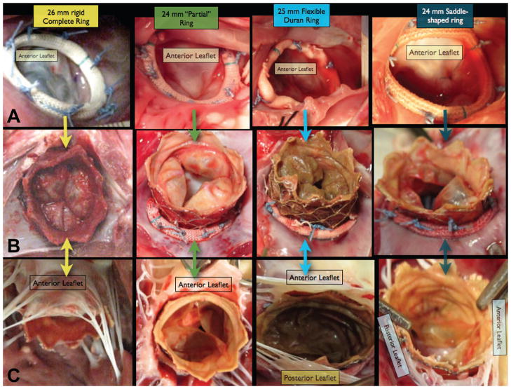
Figure 3.
Row A shows the intraoperative images following surgical Mitral annuloplasty with 4 different types of annuloplasty ring. Row B shows necropsy pictures of the atrial side of the Melody Valve-in-Ring complex. Note that there is a complete circumferential seal formed between the Melody valve and the annular tissue, even in the absence of a complete ring (column 2). Row C shows the necropsy pictures of the ventricular side of the Melody Valve-in-ring complex. In all cases, the Mitral leaflet tissue “hugs” the outside of the Melody device. Although the Melody valve leaflets appear “knarled” and “rolled”, in most cases the valves functioned well, with only one having hemodynamically significant Mitral regurgitation.

