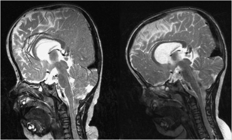
Figure 6.
Magnetic resonance images showing a reduction of tonsillar herniation and a more normal cerebrospinal fluid distribution preoperatively on the left and 3 months postoperatively on the right. (Reprinted with permission from White N, Evans M, Dover MS, et al. Posterior calvarial vault expansion using distraction osteogenesis. Child's Nervous System 25, 231–236).

