Figure 2.

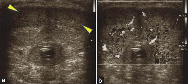
Gray-scale ultrasound images (a, b) show hypoechoic areas (yellow arrowhead), with intra-lesional vascularization on color flow Doppler examination, near the dorsal surface of the both corpora cavernosa.

Gray-scale ultrasound images (a, b) show hypoechoic areas (yellow arrowhead), with intra-lesional vascularization on color flow Doppler examination, near the dorsal surface of the both corpora cavernosa.