
Figure 3.
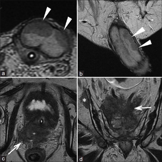
The T2-weighted images (a: axial and b: coronal) show the presence of nodules adherent to both cavernous bodies (white arrowheads). The T2- weighted images (c) and (d) show the loss of the normal architecture of the prostate due to solid tissue (recurrence of disease) which invades the region of the seminal vesicles and infiltrates the mesorectum (white arrows). Image (d) shows a bone metastasis (asterisk).