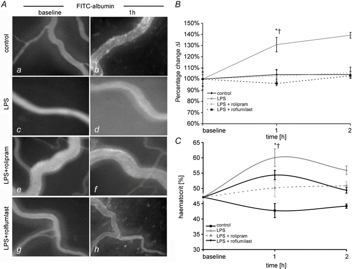
Figure 3. PD-4-Is blocked capillary leakage in postcapillary venules.

A, postcapillary mesenteric venules following i.v. application of FITC-albumin are shown under baseline conditions (a, d, g) and after 1 h (b, e, h) of experimental procedures. B, quantifications of extravasated FITC-albumin in postcapillary venules are shown. ΔI is the change in light intensity measured inside and outside the vessels. C, changes of haematocrit values in the different groups are shown. *P < 0.05 vs. control; †P < 0.05 vs. LPS+PD-4-I.
