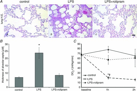
Figure 5. PD-4-Is attenuated LPS-induced lung oedema.

A, representative images of H&E stained sections of the lung demonstrate that LPS (b) induced severe lung oedema and cellular infiltration whereas these changes were not observed in the LPS+PD-4-I groups (b). Controls (a) displayed normal morphology; scale bar is 20 μm. B, quantification of thickness alveolar septa in the different experimental groups are shown. C, oxygen delivery index (DO2-I) as an overall parameter of oxygen supply was reduced in LPS-treated animals compared to the LPS+PD-4-I group whereas controls remained unchanged. *P < 0.05 vs. control; †P < 0.05 vs. LPS+PD-4-I.
