
Fig. (3).
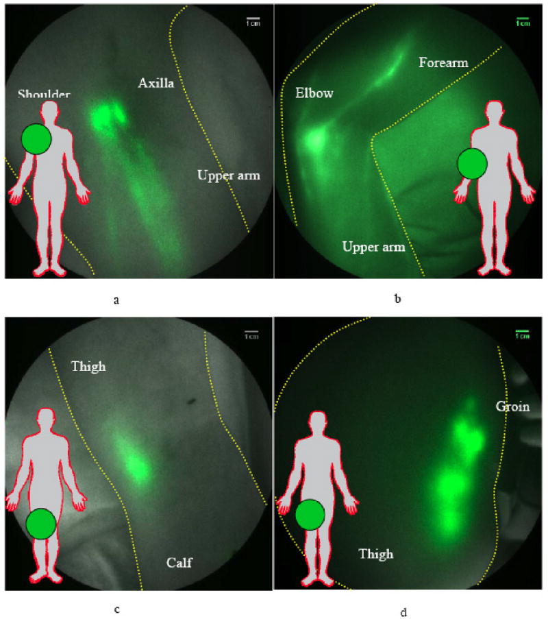
Fluorescence images of nodal regions in normal subjects: (a) three median lymphatic bundles that pool the fluorescent dye into three lymph nodes in the axilla; (b) afferent and efferent lymphatic vessels feeding and draining the fluorescent cubital lymph node in the medial forearm and elbow; (c) the popliteal lymph node in the back of the right knee (d) fluorescent signals demarking up to six superficial inguinal lymph nodes. Reproduced from Sevick-Muraca [75] with permission.